

Neurosurgery and Neurology · Jaipur Hospital
Hydrocephalus Surgery in Jaipur
When balance issues, memory changes, or headaches disrupt daily life, Jaipur Hospital provides a clear clinical evaluation and surgical guidance.
- Serving Jaipur since 1991 (35 years)
- NABH accredited
- 24×7 emergency & ICU
- Cashless: RGHS, MAA Yojana, CGHS + 22 insurers
- Operation time
- 1 to 2 hours
- Anaesthesia
- General anaesthesia
- Recovery
- Resume light activity in 1 week, avoid heavy lifting for 4 to 6 weeks
- Price
- ₹1,50,000 to ₹3,50,000typical range in India; ask us for an estimate
Fluid buildup affecting daily life
Unsteadiness, memory changes, and persistent headaches
Extra fluid builds pressure around the brain. This can lead to unsteadiness while walking, mental confusion, tiredness, or unexpected bladder issues.
-
Trouble walking and balance
Walking may feel heavy or clumsy, making regular movements slow and uncertain.
-
Headaches and nausea
Excess fluid pressure can trigger severe headaches, tiredness, and sick feelings.
-
Bladder control difficulties
Sudden urinary urgency or frequent urges to void may disrupt sleep and daily routines.
-
Cognitive changes
Slowness in thinking, forgetfulness, or mild confusion can affect independence.
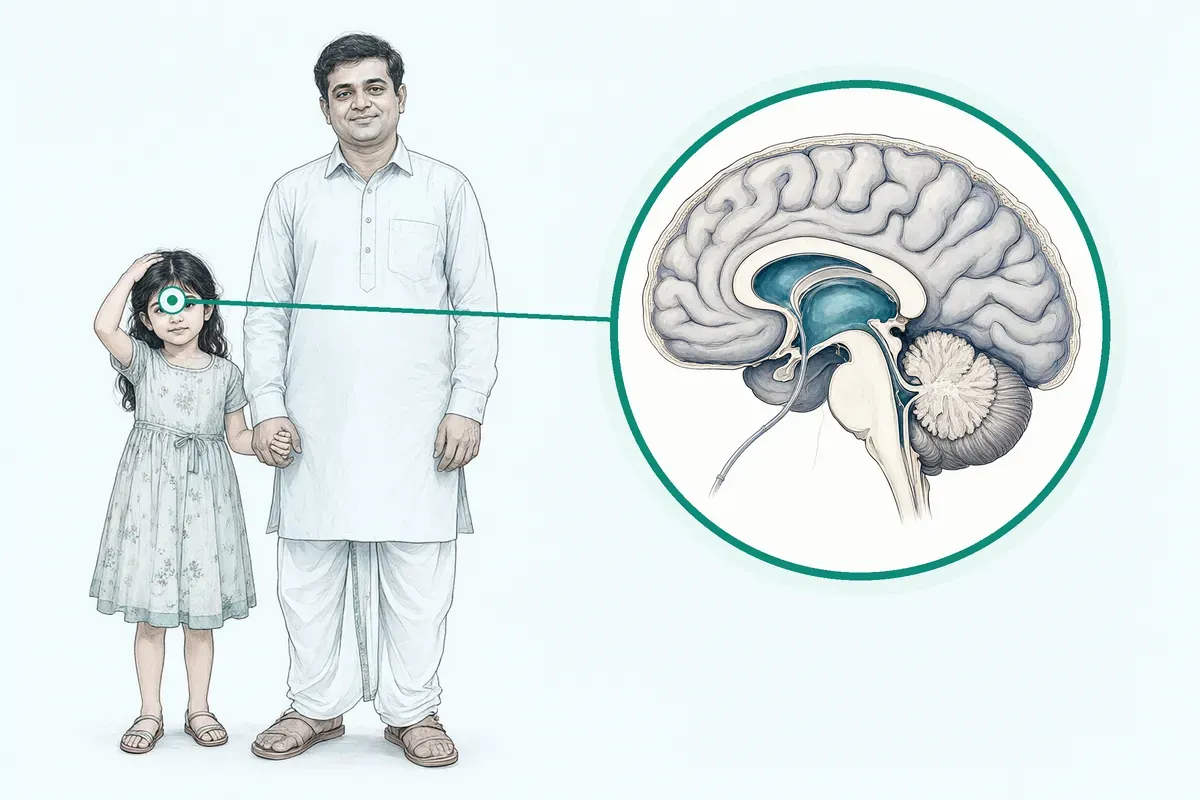
Extra cerebrospinal fluid makes the ventricles grow bigger and can raise pressure around the brain.
Why Jaipur Hospital
You may worry about the risks of brain surgery, prolonged time away from work, and treatment costs.
Your care plan
Your first steps with us
- 1 Consult our neurosurgeon in the OPD
- 2 Review your brain scans and laboratory tests
- 3 Discuss surgical options and plan care
- 4 Recover with guided medical follow-up visits
How hydrocephalus surgery is performed
Surgeries are typically performed under general anaesthesia to divert excess fluid. The operation usually takes 1 to 2 hours.
-
Ventriculoperitoneal shunt
A standard option your surgeon will discuss to divert excess fluid into the abdomen using a catheter and valve.
-
Endoscopic third ventriculostomy
An option your surgeon will discuss for obstructive hydrocephalus that creates a bypass opening and avoids hardware.
-
Ventriculoatrial shunt
An alternative diversion your surgeon will discuss when the peritoneal route is not clinically feasible.
-
Shunt revision
A procedure your surgeon will discuss if an existing shunt develops malfunction, infection, or blockage.
Why patients trust us
- 1991 Serving Jaipur for 35 years
- NABH Accredited hospital
- 1 Neurosurgery and Neurology consultants
- 24×7 Emergency, ICU and ambulance
- 25 Cashless schemes and insurers
Clinical findings on shunt procedures
-
82 %
Studies published over a recent 5-year period showed improvement following shunt insertion in evaluated patients.
-
8.2 %
Studies reported a combined common complication rate of 8.2 percent following shunt insertion.
-
1 to 2 days
Most patients in study protocols stayed in the hospital for 1 to 2 days after VP shunt placement.
If it is left untreated
Leaving hydrocephalus untreated can allow ventricles to enlarge further, worsening walking difficulty, urinary incontinence, and cognitive decline over time.
Life after surgical treatment
With excess fluid managed, most patients gradually resume familiar routines under medical guidance.
-
Stepwise daily recovery
Patients usually resume light activity in 1 week while avoiding heavy lifting for 4 to 6 weeks.
-
Hospital discharge
A hospital stay typically lasts 3 to 5 nights before continuing recuperation at home.
-
Ongoing shunt monitoring
Without problems, a shunt device may remain safely in place for years with regular reviews.

Frequently asked questions
Questions patients ask about hydrocephalus surgery
Which problems are assessed under hydrocephalus and CSF disorders?
In short
At Jaipur Hospital, our neurosurgery team assesses conditions where cerebrospinal fluid builds up and raises pressure inside the skull. This includes congenital hydrocephalus present from birth, normal pressure hydrocephalus in older adults, and obstructive hydrocephalus caused by tumours or scarring. Post-haemorrhagic and post-infectious hydrocephalus are also evaluated.
In detail
Hydrocephalus develops when the production, flow, or absorption of cerebrospinal fluid is disturbed. In infants, enlarged ventricles may be seen on prenatal scans or after birth with a rapidly growing head.
In adults, normal pressure hydrocephalus classically presents with the triad of gait disturbance, cognitive decline, and urinary incontinence. Obstructive forms occur when a tumour, cyst, or aqueductal stenosis blocks the ventricular pathways. Post-haemorrhagic hydrocephalus follows intraventricular bleeding, while post-infectious hydrocephalus follows meningitis. Each type has different causes and progression, so the surgical team tailors the assessment to the underlying mechanism.
A common belief
Many people think hydrocephalus only affects babies, but normal pressure hydrocephalus is common after age 60 and is often mistaken for dementia or Parkinson disease.
What actually happens
Correct diagnosis changes the treatment path completely.
Your next step at Jaipur Hospital
You will need a clinical examination and brain imaging, usually MRI or CT, to confirm ventricle size and look for a cause. The surgeon reviews the images, checks your symptoms, and decides whether shunt surgery or endoscopic third ventriculostomy is appropriate.
How will the doctor decide which tests I need for hydrocephalus?
In short
At Jaipur Hospital, the neurosurgeon selects tests based on your age, symptoms, and suspected type of hydrocephalus. MRI brain with CSF flow studies is the standard first investigation. CT brain is used when MRI is contraindicated or for quick ventricle size assessment. Additional tests may include lumbar puncture with pressure measurement or tap test for normal pressure hydrocephalus.
In detail
MRI shows ventricle size, periventricular oedema, aqueduct patency, and any tumour or cyst causing obstruction. CSF flow sequences assess aqueductal stroke volume.
In normal pressure hydrocephalus, a diagnostic lumbar puncture measures opening pressure and a tap test checks whether removing 30 to 50 ml of CSF improves gait. CT is faster and useful in emergencies or when a shunt series is needed to check catheter position.
Blood tests include complete blood count, coagulation profile, renal and liver function, and infection markers for anaesthesia clearance. The surgeon orders only the tests that change management, avoiding unnecessary radiation or invasive procedures.
A common belief
Some centres order every scan routinely.
What actually happens
Our team orders only what changes the plan — MRI for anatomy and flow, tap test for NPH diagnosis, CT only when needed. This avoids extra cost and radiation.
Your next step at Jaipur Hospital
Expect MRI brain as the primary test. If normal pressure hydrocephalus is suspected, a tap test may be done as a day-case. Blood tests are drawn before surgery. Results are usually available within 24 to 48 hours.
How soon should I expect improvement after hydrocephalus surgery?
In short
After hydrocephalus surgery at Jaipur Hospital, some symptoms such as headache may improve within days, while gait and cognitive changes often take several weeks to show measurable progress. The speed of recovery varies with the underlying cause, age, and any co-existing medical conditions.
In detail
Evidence indicates that headache and nausea frequently ease soon after cerebrospinal fluid diversion. Walking difficulty and memory problems typically improve gradually over weeks to months as the brain adjusts to normal pressure.
In studies of idiopathic normal pressure hydrocephalus, favourable outcome defined as a one-point gain on the modified Rankin scale was seen at three months in a majority of treated patients.
However, the presence of comorbidities such as stroke, small vessel disease, or Alzheimer disease can limit the extent of recovery. Children with congenital hydrocephalus may need regular follow-ups to monitor growth and shunt function.
Shunt valves that are programmable allow pressure adjustments after surgery, which can fine-tune drainage and optimise symptom control over time.
A common belief
A common belief is that all symptoms vanish immediately.
What actually happens
In practice, the brain needs time to adapt to normal pressure. Comorbidities like stroke or dementia can slow or limit recovery, and programmable shunts may need a few adjustments to find the right setting.
Your next step at Jaipur Hospital
Plan for a hospital stay of three to five nights. Light activity resumes in about one week, but avoid heavy lifting for four to six weeks.
Attend all scheduled follow-ups so the surgical team can assess shunt function or endoscopic patency and adjust settings if needed. Keep a symptom diary to track changes in walking, memory, and urinary habits.
How does the term "cerebrospinal fluid" relate to hydrocephalus and CSF disorders?
In short
Cerebrospinal fluid (CSF) is the clear fluid that cushions the brain and spinal cord, brings nutrients, and removes waste. In hydrocephalus, extra CSF builds up in the brain ventricles, raising pressure and causing symptoms. Surgery at Jaipur Hospital restores normal CSF flow or drainage.
In detail
CSF is produced continuously inside the ventricles. Normally it flows through narrow pathways, bathes the brain and spinal cord, and is absorbed into the bloodstream. When production exceeds absorption or a blockage stops the flow, the ventricles enlarge. This is hydrocephalus.
The extra fluid stretches the ventricles and presses on surrounding brain tissue, leading to headache, nausea, gait disturbance, cognitive decline, and urinary incontinence. A VP shunt diverts CSF from the ventricles to the peritoneal cavity in the abdomen where the body absorbs it.
An endoscopic third ventriculostomy creates a new opening in the floor of the third ventricle so CSF can bypass the blockage and reach the absorption sites. Both treatments aim to rebalance CSF dynamics and lower intracranial pressure.
A common belief
A common misunderstanding is that CSF is just water.
What actually happens
It is a specialised fluid with nutrients and waste-removal functions. Simply removing fluid without addressing the flow problem can lead to over-drainage. That is why programmable valves and endoscopic techniques are used to fine-tune the balance.
Your next step at Jaipur Hospital
If you have symptoms suggesting hydrocephalus, the neurosurgery team will order CT or MRI scans to visualise the ventricles and CSF spaces. They will explain whether a shunt or endoscopic procedure is appropriate for your type of CSF disorder.
How will pressure or fluid-flow problems be confirmed before hydrocephalus surgery?
In short
Before hydrocephalus surgery at Jaipur Hospital, pressure and fluid-flow problems are confirmed through clinical assessment, imaging, and sometimes direct pressure measurement. MRI or CT scans show ventricle size and flow patterns, while a lumbar puncture with pressure monitoring can document elevated CSF pressure and test symptom response to fluid removal.
In detail
The neurosurgeon reviews your history for headache, gait disturbance, cognitive changes, and urinary symptoms. Imaging reveals ventriculomegaly and may show disproportionately enlarged subarachnoid spaces, a feature associated with idiopathic normal pressure hydrocephalus.
In some centres, a tap test removes 30 to 50 ml of CSF via lumbar puncture and observes for gait or memory improvement over hours to days. A positive response supports shunt benefit.
For obstructive hydrocephalus, cine MRI or phase-contrast sequences can visualise aqueductal flow voids. Intracranial pressure monitoring via a bolt or sensor may be used in complex cases. Blood tests, coagulation profiles, and anaesthesia clearance are completed before any procedure.
A common belief
A scan alone does not always confirm the need for surgery.
What actually happens
Some patients have large ventricles but few symptoms. The tap test and pressure monitoring help separate true hydrocephalus from ventriculomegaly that does not need intervention.
Your next step at Jaipur Hospital
Expect a pre-operative workup spanning a few days: imaging, blood tests, and anaesthesia review. If a tap test is planned, it may require a short hospital stay for observation.
The surgical team will explain each test and how the results guide the choice between shunt insertion and endoscopic third ventriculostomy.
How can I decide whether to seek advice about ventriculoperitoneal shunt surgery?
In short
You should seek advice about ventriculoperitoneal shunt surgery when imaging shows enlarged ventricles and you have symptoms such as persistent headache, nausea, difficulty walking, memory problems, or urinary incontinence that affect daily life. Jaipur Hospital evaluates each case with a neurosurgeon to confirm the indication.
In detail
The decision rests on matching clinical symptoms to radiological findings. Evidence states that shunt insertion is recommended when there is a favourable risk‑to‑benefit ratio, and that symptomatic improvement can be achieved by surgical placement of a suitable CSF shunt device.
In idiopathic normal pressure hydrocephalus, studies report positive improvement in an average of 71 percent of patients, with more recent series showing 82 percent improvement. However, comorbidities such as stroke, dementia, or significant cardiac disease lower the chance of a fully favourable outcome.
The neurosurgeon reviews MRI or CT scans, performs a clinical assessment, and may arrange a tap test or lumbar drain trial to predict response before offering surgery.
A common belief
Some patients wait until they fall or become dependent, but early evaluation improves the chance of meaningful recovery.
What actually happens
Comorbidities like stroke or dementia reduce the benefit, so the surgeon weighs each factor before recommending surgery.
Your next step at Jaipur Hospital
If you or your relative have the described symptom cluster, request a neurosurgery referral. The team at Jaipur Hospital will arrange imaging, clinical scoring, and, if appropriate, a tap test within a few days. Surgery typically takes one to two hours under general anaesthesia with a three‑to‑five‑night stay.
What changes in strength, sensation, speech, vision, or bladder control need urgent assessment?
In short
Sudden weakness, numbness, slurred speech, vision loss, new seizures, or loss of bladder or bowel control need urgent assessment after hydrocephalus surgery. Jaipur Hospital provides 24-hour emergency care for these neurological changes.
In detail
These signs may indicate shunt malfunction, intracranial haemorrhage, seizure, or new neurological injury. Evidence shows serious adverse event risk of 11% in iNPH. The neurosurgical team evaluates with urgent imaging and clinical examination. Post-operative monitoring includes regular neurological checks for strength, sensation, and cognition. Any acute change warrants immediate hospital review.
- Evidence shows serious adverse event risk of 11% in iNPH.
A common belief
People sometimes hope these will pass, but they can mean the shunt has failed or there is bleeding.
What actually happens
Delay can cause long-term damage.
Your next step at Jaipur Hospital
Go to the emergency department or call an ambulance if these symptoms appear suddenly. Do not wait for an outpatient appointment. The team will investigate and treat the cause promptly.
Which symptoms should prompt me to see a specialist?
In short
At Jaipur Hospital, you should see a neurosurgery specialist if you notice persistent headache, nausea, vomiting, blurred vision, difficulty walking, memory problems, or urinary incontinence. In infants, a rapidly increasing head size, bulging fontanelle, sunset eyes, or irritability are warning signs. These symptoms suggest raised intracranial pressure or disturbed CSF flow.
In detail
Headache is the most common symptom of hydrocephalus and often worsens in the morning or with lying down. Nausea and vomiting may accompany the headache. Vision changes such as blurred or double vision occur when pressure affects the optic nerves.
In normal pressure hydrocephalus, the classic triad is gait disturbance with a magnetic or shuffling walk, cognitive slowing, and urinary urgency or incontinence.
Infants cannot describe headache, so a tense or bulging fontanelle, prominent scalp veins, and the sunset sign (eyes fixed downward) are key clues. Any new neurological deficit or change in consciousness also warrants urgent review.
A common belief
People often dismiss morning headaches or frequent falls in the elderly as normal ageing.
What actually happens
In normal pressure hydrocephalus, these are treatable signs. Missing them delays surgery that can improve walking and memory.
Your next step at Jaipur Hospital
Keep a symptom diary noting when headaches occur, any vision changes, and walking or memory difficulties. Bring previous imaging reports if you have them. The specialist will examine you, order MRI or CT if needed, and discuss whether surgery is indicated.
How will the results change my care plan for hydrocephalus?
In short
At Jaipur Hospital, imaging and test results directly guide whether you need surgery, which procedure suits you, and how urgent it is. Large ventricles with obstruction favour endoscopic third ventriculostomy. Normal pressure hydrocephalus with a positive tap test supports shunt insertion. Stable, asymptomatic ventriculomegaly may only need monitoring.
In detail
MRI shows the site of obstruction. If the aqueduct is blocked but the floor of the third ventricle is thin, ETV can bypass the blockage without hardware.
If the obstruction is at the foramina of Luschka and Magendie, or if previous ETV failed, a ventriculoperitoneal shunt with a programmable valve is preferred. In normal pressure hydrocephalus, a clear gait improvement after lumbar tap test predicts shunt response.
If the tap test is negative, other causes of gait and memory problems are investigated. Shunt series X-rays or CT check catheter position if malfunction is suspected. Blood results determine anaesthesia fitness and infection risk. Each result narrows the options to the safest, most effective approach for your anatomy.
A common belief
Patients often assume all hydrocephalus gets a shunt.
What actually happens
In fact, ETV avoids lifelong hardware in suitable anatomy. A negative tap test in NPH redirects care to other diagnoses. The right test prevents the wrong surgery.
Your next step at Jaipur Hospital
After tests, the surgeon explains the findings, recommends shunt or ETV, and discusses timing. Surgery is usually scheduled within one to two weeks if indicated. If monitoring is chosen, follow-up imaging is set at three to six months.
What should I do if symptoms persist or worsen after hydrocephalus surgery?
In short
Contact your neurosurgeon or the emergency department at Jaipur Hospital if headaches return, fever develops, the wound becomes red or leaks fluid, vomiting starts, or you notice new swelling along the shunt track.
In detail
Shunt malfunction, infection or blockage can occur weeks to years after insertion. Signs include recurrence of original symptoms such as gait disturbance, confusion or incontinence, plus wound changes, abdominal pain or a palpable lump along the catheter.
In studies, the combined complication rate after shunt insertion is around eight percent, with serious adverse events in about eleven percent of idiopathic normal pressure hydrocephalus cases. Endoscopic third ventriculostomy avoids hardware but can fail if the stoma closes. Prompt imaging and clinical review allow early revision if needed.
A common belief
Some patients assume a shunt lasts forever without trouble.
What actually happens
In reality, blockage or infection can happen at any time, so knowing the red flags is essential.
Your next step at Jaipur Hospital
Do not wait for the next routine visit if warning signs appear. Keep the hospital numbers handy and seek care the same day. The team can arrange urgent CT or MRI, shunt tap or valve reprogramming.
FAQ library
Questions and answers about hydrocephalus surgery
When you contact us
Schedule your neurosurgery consultation in Jaipur
Contact our team to book an OPD assessment and review your brain scans.
- A consultation with a neurosurgeon in the OPD
- Your scans and reports reviewed, with tests done in-house if you need them (laboratory, X-ray and imaging)
- Your options explained in plain words, including whether you need surgery at all
- A cost estimate before you decide
- Help with cashless treatment under RGHS, MAA Yojana, CGHS and empanelled insurers and TPAs
- Follow-up visits after your treatment
Overview
Hydrocephalus Surgery: what to know
What is hydrocephalus and how does it develop
Cerebrospinal fluid, or CSF, is a clear fluid that cushions and protects your brain and spinal cord. This fluid also carries essential nutrients throughout your nervous system and removes cellular waste. Your brain normally produces and absorbs this fluid continuously to maintain a healthy balance.
Hydrocephalus happens when excess cerebrospinal fluid builds up inside the natural cavities of your brain, known as ventricles. As extra fluid accumulates, it causes these ventricles to enlarge. This enlargement raises pressure inside your skull.
This elevated pressure presses against sensitive brain tissue. Over time, the pressure interferes with normal brain function and can make daily activities difficult. In elderly individuals, a specific form called idiopathic normal pressure hydrocephalus can develop, characterised by enlarged ventricles along with walking problems, memory decline, and bladder control issues.
Symptoms of hydrocephalus and when to seek emergency care
- Sudden severe headache Begins suddenly or worsens rapidly
- Severe trouble breathing Call emergency services immediately
- Seizures or jerking movements Shaking or jerking of the body
- Persistent vomiting Does not stop
- Fall with head injury Hit your head during a fall
- High fever or sudden confusion Sudden worsening of confusion
The most common symptom of hydrocephalus is a headache. You may also feel unusual tiredness or experience nausea. As fluid pressure builds within the brain, it can lead to confusion and difficulty with walking or balance.
Changes in bladder control are also frequent with this condition. Many people experience sudden urinary urgency, a need to urinate frequently, or urinary incontinence. In studies of adults with idiopathic normal pressure hydrocephalus, urinary difficulties were reported in 55% to 79% of individuals. These bladder issues often occur because of sudden, involuntary bladder contractions.
Certain warning signs require urgent medical care the same day. You or your attendant should seek immediate emergency medical care if you experience any of the following:
- Severe headache that begins suddenly or worsens rapidly
- Severe trouble breathing
- Shaking, jerking movements, or seizures in your body
- Persistent vomiting
- A fall where you hit your head
- A high fever or sudden worsening in your confusion
Causes of hydrocephalus and who is at higher risk
- 175 per 100,000 General population
- 400 per 100,000 Aged 80 years and older
Hydrocephalus can arise from several different medical factors. Extra cerebrospinal fluid may be present when a person is born, known as congenital hydrocephalus. In other cases, extra fluid builds up later in life due to an obstruction, such as a brain tumour that blocks normal fluid pathways.
It can also occur when other central nervous system diseases coexist, including:
- Stroke
- Small vessel disease
- Parkinson's disease
- Dementia
- Spinal conditions
Age is a major risk factor for certain forms of hydrocephalus. In a worldwide 2018 meta-analysis, the mean prevalence of idiopathic normal pressure hydrocephalus was estimated at 175 per 100,000 people in the general population, rising to 400 per 100,000 among individuals aged 80 years and older.
In two Swedish surveys using head CT scans, prevalence rates were 8.9% and 5.9% in people aged 80 years and above.
Other health conditions increase the likelihood of developing complications like bladder control issues. Research indicates that older age, longer illness duration, reduced functional mobility, diabetes, and the use of cholinesterase inhibitor medicines are associated with higher rates of urinary incontinence. People who have had a prior shunt infection or shunt blockage also carry a higher risk of recurrent fluid buildup.
How hydrocephalus is diagnosed: examinations and tests
- Clinical evaluation Neurosurgeon checks walking, memory, bladder
- Brain imaging MRI or CT scan for enlarged ventricles
- Spinal MRI if needed For spinal cord compression symptoms
- Bladder assessment History, infection check, bladder diary
- Lumbar tap test Fluid removed to test walking or bladder improvement
Diagnosing hydrocephalus begins at Jaipur Hospital with a clinical evaluation by a neurosurgeon. Your doctor conducts a physical examination, reviews your medical history, and checks for difficulties with walking, memory, and bladder function.
Brain imaging is essential to confirm the diagnosis. Your surgeon will arrange an MRI or CT scan of your brain to look for enlarged ventricles and examine fluid spaces. In patients being evaluated for idiopathic normal pressure hydrocephalus, scans often reveal disproportionately enlarged subarachnoid spaces and tightness near the top of the brain.
If you also have symptoms suggesting spinal cord compression, a spinal MRI is performed to check your neck and back.
Your medical team may also evaluate your bladder health. This includes taking a detailed history of how often you urinate, checking for urinary tract infections, and assessing whether your bladder empties fully. You may be asked to keep a bladder diary for a few days to record fluid intake, urination frequency, and leakage episodes.
A lumbar tap test may be performed, where a small amount of fluid is removed from the lower back to observe whether your walking or bladder control temporarily improves.
Treatment options and when hydrocephalus surgery is advised
Hydrocephalus involves physical fluid buildup inside the skull, so medications alone cannot correct structural fluid blockage or enlarged ventricles. Non-surgical care focuses on identifying and managing coexisting chronic medical conditions. Our medical team treats underlying health problems such as high blood pressure, diabetes, heart conditions, or urinary infections to protect your overall health.
When patients have other conditions, such as progressive cervical spinal cord compression or severe spinal canal stenosis, those spinal issues may need treatment before or alongside hydrocephalus care.
Surgery is advised when hydrocephalus symptoms significantly interfere with your daily life and tests show that relieving brain pressure may help. Your neurosurgeon recommends surgical diversion when the potential benefits outweigh the surgical risks.
Surgery is indicated for infants born with structural fluid blockage, individuals with recurrent fluid buildup or shunt malfunction, and eligible adults showing progressive gait problems or cognitive decline.
In studies of patients undergoing shunt surgery, positive improvement was reported in an average of 71% of cases overall, and in 82% of patients in studies published in recent years. However, shunt surgery carries an 11% risk of serious adverse events in idiopathic normal pressure hydrocephalus, and recent studies report an 8.2% combined common complication rate. Your surgeon will discuss your imaging, symptoms, and health conditions to advise whether surgery is appropriate for you.
Preparing for hydrocephalus surgery
Do
- Tell your doctor about all medicines Include blood thinners like aspirin
- Inform if pregnant or planning pregnancy So care can be monitored
- Arrange a family attendant To accompany and assist after discharge
Avoid
- Avoid alcohol and heavy meals Night before operation
Preparation for hydrocephalus surgery involves several steps to help the procedure proceed smoothly. Your preparation begins with a comprehensive evaluation by your neurosurgeon. Your neurosurgeon specialises in surgery of the brain and nervous system. This assessment may include a physical examination, a review of your medical history, and brain imaging studies such as CT scans or MRI scans. You may also undergo blood tests to check coagulation profiles, screen for infections, and complete other routine assessments.
Medical clearance for general anaesthesia requires addressing underlying conditions such as cardiac disease or uncontrolled high blood pressure. An anaesthesiologist creates the anaesthesia plan and monitors you during the operation.
The anaesthesiology team may ask you about:
- Your age
- Height
- Weight
- Prior surgeries
- Any family or personal history of anaesthesia problems
You must talk to your doctor about your current medicines, including blood thinners such as aspirin, so they can tell you when to pause or restart them. If you are pregnant or planning pregnancy, inform your healthcare provider so they can monitor your care. If a patient has spina bifida, our surgical team must be told because of the risk of latex allergy.
In the days before surgery, eat a balanced diet with vegetables, fruits, and lean proteins. Avoid alcohol and heavy meals the night before your operation. You are typically instructed to stop eating and drinking for a set period before the procedure, usually starting the night before surgery.
You may be asked to bathe with an antiseptic soap on the morning of surgery or the night before to reduce skin infection risks.
Because the operation is done under general anaesthesia, arrange for a family attendant to accompany you, stay with you, and assist you after discharge. Bring a written list of all your current medicines and your prior test reports to the hospital. You should discuss any remaining questions or concerns with our surgical team before your procedure.
How hydrocephalus surgery is done
Hydrocephalus surgery diverts excess fluid away from the brain to relieve pressure. Your surgeon will advise which surgical technique suits your condition.
Several surgical techniques exist to manage cerebrospinal fluid:
- Ventriculoperitoneal shunt: Standard CSF diversion with programmable valve. A catheter is placed into a brain ventricle and connected to a valve. A second catheter runs under the skin from behind the ear, down the neck, and into the belly, where the body absorbs the fluid.
- Endoscopic third ventriculostomy: ETV for obstructive hydrocephalus, avoids shunt hardware.
- Ventriculoatrial shunt: Alternative diversion when peritoneal route not feasible. It passes the lower catheter under the skin into the right atrium of the heart, where fluid enters the bloodstream.
- Shunt revision: Revision for shunt malfunction, infection or blockage.
When a shunt is chosen, different valve designs are available based on your clinical needs. Programmable shunts allow your doctor to adjust valve pressure settings after surgery without another operation. Fixed pressure shunts have a set pressure setting that cannot be changed after placement. Shunts may also include antisiphon devices to prevent over-drainage when you change postures.
In select situations, surgeons may consider dual-lumen shunts or systems with integrated sensors. Other pathways include ventriculopleural shunts into the chest cavity or lumboperitoneal shunts from the lower spine to the belly.
On the day of your operation, you will check in at Jaipur Hospital and be received in the pre-operative area. A nurse will check your vital signs, confirm your details, and place an intravenous line for fluids and medicines. Hydrocephalus surgery is typically performed under general anaesthesia, keeping you asleep and comfortable throughout the procedure. Operation time is typically 1 to 2 hours, and a standard shunt procedure takes about 1 hour.
For a ventriculoperitoneal shunt, your surgeon makes small incisions: typically on the scalp, behind the ear or neck, and on the abdomen. The catheter is guided into the ventricle, the valve is secured, and the lower catheter is passed through the fatty tissue under the skin into the abdominal cavity. The surgeon tests fluid flow through the system before closing.
The entire shunt system stays beneath the skin. Incisions are closed using stitches or surgical staples, and sterile dressings are applied.
Risks and complications of hydrocephalus surgery
- 8.2% Combined common complications
- 11% Serious adverse events in iNPH
- 1% Average mortality after shunt insertion
- 0.2% Mortality in recent surgical studies
Every surgical procedure carries potential risks. In recent medical studies, a combined common complications rate of 8.2% was reported following shunt insertion. In studies evaluating idiopathic normal pressure hydrocephalus, the risk of serious adverse events was reported at 11%. Because of these risks, surgeons carefully evaluate whether the expected benefits outweigh the risks before recommending an operation.
Published studies report an average mortality rate of 1% following shunt insertion. In more recent surgical studies, this mortality rate was reported at 0.2%.
Complications can include:
- Shunt malfunction
- Catheter blockage
- Infection
If a shunt gets infected or stops working, your surgeon may need to adjust, clear, or replace the hardware. Other conditions can affect how smoothly recovery goes. In elderly patients, co-existing health problems such as diabetes, high blood pressure, stroke, or heart disease can influence surgical risks and overall progress.
Incisions can occasionally become tender, open up, or develop fluid collections. In some cases, fluid drainage may require minor adjustments of a programmable valve in the OPD to achieve balanced drainage.
Recovery and aftercare following surgery
- Hospital stay Typically 3 to 5 nights with attendant support
- Light activity Resume in 1 week
- Desk work Return in less than 1 week for most people
- Heavy lifting Avoid for 4 to 6 weeks
- Wound care Keep dressings dry for 5 days, then shower after surgeon approval
- Stitches or staples removal During OPD follow-up visit
Immediately after surgery, you will be cared for in the recovery room. A nurse will monitor your pulse, blood pressure, temperature, and breathing. The nurse will ask you questions to evaluate brain function and ask you to move your arms, legs, fingers, and toes. You will then be transferred to your hospital room.
A hospital stay is typically 3 to 5 nights. Your attendant can stay to assist you with moving around and daily needs.
During your hospital stay, mild soreness around incisions in the neck, head, or abdomen is normal, but severe pain is uncommon. You may feel a raised bump on your head where the valve rests under the scalp. You might also feel fluid moving under the scalp for a short time.
Take all prescribed pain medicines and antibiotics exactly as directed by your doctor. If pain medicine causes an upset stomach, take it with food or ask your doctor about changing it. Do not combine pain medicines unless your doctor explicitly approves. You can buy your medicines from the in-house pharmacy or a local chemist.
At home, resume light activity in 1 week, and avoid heavy lifting for 4 to 6 weeks. Most people return to routine desk work in less than 1 week. Avoid contact sports or any activity that risks a blow to your head or neck.
Do not press or rub the shunt valve on your head, though resting your head on that side is safe. For the first 5 days, keep your dressings dry and do not shower. Once your surgeon approves bandage removal, you may shower, but avoid tub baths and swimming until stitches or staples are removed.
If needed, your surgeon will remove your stitches or staples during an OPD follow-up visit. Do not drive or drink alcohol while on prescription pain medications. Check with your doctor before booking air travel.
Eat light meals at first, gradually returning to normal food. The abdominal catheter does not alter normal food digestion. If your stomach is unsettled, choose bland foods such as plain rice, curd, toast, or broiled chicken.
Constipation can occur after surgery; avoid straining and consult your doctor about a mild laxative if you have no bowel movement for a few days. For elderly patients, attendants should assist with walking, monitor wound healing, and maintain medicine schedules.
Contact your doctor or visit our OPD promptly if you experience any of these signs:
- Fever, or redness and red streaks spreading from an incision
- Pus or clear fluid draining from the incision, or edges of the wound opening
- A new bump on your head 3 to 5 days after surgery, or a bump enlarging after 2 weeks
- New, worsening, or persistent headaches, or frequent vomiting
- New or worsening abdominal pain
- If you fall and hit your head
Seek immediate emergency medical care if you develop severe trouble breathing, seizures with body jerking or shaking, or a sudden, severe headache.
Expected results and study outcomes
- 71% Average improvement across 64 studies
- 82% Improvement in recent 5-year studies
- 96% Subjective symptom improvement at 6 months
The primary goal of hydrocephalus surgery is to lower raised brain pressure and improve symptoms such as trouble walking, headache, tiredness, and confusion. When shunts function without complications, they can remain in place for years.
Clinical studies provide data on outcomes across groups of patients. In a review of 64 clinical studies including 3,063 patients, positive improvement following shunt surgery was reported in an average of 71% of patients. Among studies published in the subsequent 5 years, an 82% improvement rate was reported after shunt insertion.
In one clinical trial comparing immediate surgery against delayed treatment in 93 patients, 65% of patients in the immediate surgery group showed functional improvement on the modified Rankin scale at 3 months, compared to 5% in the delayed group.
In another study evaluating normal pressure hydrocephalus, researchers reported a 96% chance of subjective symptom improvement and an 83% chance of improvement on a timed walking test at 6 months.
Recovery timelines vary between individuals. Some patients notice improvement in certain symptoms right away, while other symptoms take several weeks to show change. Long-term outcomes depend on individual factors, including overall health and the presence of underlying medical conditions such as stroke, Parkinson's disease, or vascular disease. Attending all scheduled follow-up visits allows your surgeon to check shunt function and adjust valve settings if required.
Hydrocephalus surgery cost and insurance in Jaipur
The cost for hydrocephalus surgery typically ranges from ₹1,50,000 to ₹3,50,000 in India. In published medical cost surveys, general VP shunt expenses in India typically range from ₹1,00,000 to ₹2,50,000. You can ask Jaipur Hospital for an estimate tailored to your clinical requirements.
Several factors influence the final cost of your treatment:
- The type of surgery chosen, such as an endoscopic third ventriculostomy or a shunt procedure
- The specific shunt hardware advised by your surgeon, including programmable valves or antisiphon mechanisms
- The duration of your hospital stay and room category chosen beyond the typical 3 to 5 nights
- Pre-operative investigations, blood tests, and MRI or CT scans
- Post-operative monitoring and medicines prescribed from the pharmacy
- Any management required for co-existing medical conditions during admission
Jaipur Hospital is an established, NABH-accredited multispeciality hospital in Jaipur. Patients can utilise government health schemes and coverage options accepted by the hospital, including Rajasthan Government Health Scheme (RGHS), MAA Yojana, and Central Government Health Scheme (CGHS).
The hospital also works with a panel of private health insurance companies and Third Party Administrators (TPAs) to facilitate cashless claims and reimbursement processes.