

Neurosurgery and Neurology · Jaipur Hospital
Neuro Diagnostics and Surgical Planning in Jaipur
You need clear imaging and a safe surgical plan. We provide advanced diagnostics and expert guidance.
- Serving Jaipur since 1991 (35 years)
- NABH accredited
- 24×7 emergency & ICU
- Cashless: RGHS, MAA Yojana, CGHS + 22 insurers
- Session length
- 30 to 90 minutes per test
- Anaesthesia
- Not needed or Local anaesthesia
- Recovery
- Immediate for most tests
- Price
- ₹3,000 to ₹25,000 per studytypical range in India; ask us for an estimate
Unclear scans delay safe surgery
You need to know exactly where the problem lies before surgery
Standard scans may miss critical nerve pathways. Without precise mapping, surgery carries higher risk to speech, movement or sensation. You want confidence that your surgeon sees the full picture.
-
Vague imaging results
Routine MRI may not show white matter tracts clearly, leaving the surgeon unsure of safe corridors.
-
Fear of deficits
You worry about losing speech, strength or sensation after an operation near vital pathways.
-
Multiple opinions
Conflicting advice from different doctors makes it hard to choose the right plan.
-
Seizure focus unknown
Long-term EEG monitoring is needed to pinpoint where seizures start before considering surgery.
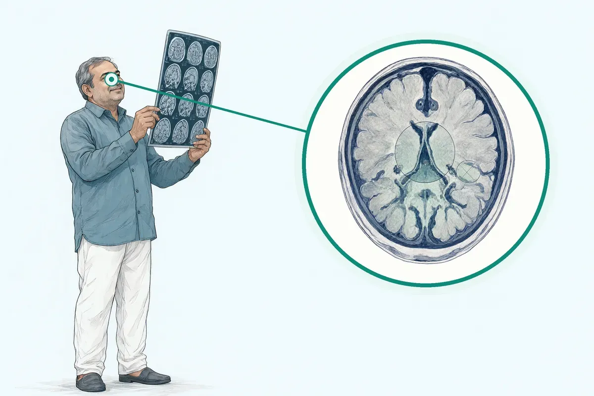
Diffusion MRI tractography enables non-invasive reconstruction of the brain's white matter connections at macro scale.
Why Jaipur Hospital
You are anxious about the operation and whether you will recover your speech, movement or independence.
Your care plan
Your first steps with us
- 1 Call or visit OPD for neurosurgery consultation
- 2 Complete advanced imaging and neurophysiology tests in-house
- 3 Review surgical plan with your surgeon
- 4 Undergo surgery with navigation and monitoring, then recover
How the assessment and planning are done
You attend as an outpatient or day-care patient. Tests take 30 to 90 minutes each, usually without anaesthesia. Your surgeon reviews all data to build a precise operative plan.
-
3T MRI with Tractography
Maps white matter tracts so your surgeon can see the relationship between a lesion and critical pathways for speech and movement.
-
Intraoperative Neuro Monitoring
Provides real-time alerts on nerve function during surgery, helping the team avoid injury to motor and sensory pathways.
-
Stereotactic Navigation
Uses preoperative imaging like GPS to guide instruments accurately during biopsy or resection.
-
Video EEG Telemetry
Records brain activity over hours or days to locate the seizure focus for epilepsy surgery planning.
Why patients trust us
- 1991 Serving Jaipur for 35 years
- NABH Accredited hospital
- 1 Neurosurgery and Neurology consultants
- 24×7 Emergency, ICU and ambulance
- 25 Cashless schemes and insurers
What research shows about advanced neuro diagnostics
-
11.93% improvement in tracking accuracy
Topology-informed pruning improved deterministic tractography accuracy in single-shell acquisitions compared to uncorrected methods.
-
Significant correlation between structural and functional connectivity
Studies show stronger structural connections correspond to stronger functional connectivity across multiple tractography approaches.
-
Reduced subcortical stimulations needed
Combining tractography with neuronavigation decreased the number of intraoperative stimulations, making surgery quicker especially for awake patients.
If it is left untreated
Without precise mapping, surgery near critical pathways carries a higher risk of new neurological deficits such as weakness or speech difficulty. Brain shift during surgery can invalidate preoperative registration if not accounted for.
Life after your diagnostic workup and surgery
Most people return to daily routines with a clear understanding of their condition and a plan tailored to preserve function.
-
Clear answers
You receive a detailed report showing the exact location of the problem relative to vital pathways.
-
Informed choices
Your surgeon explains the options, risks and expected recovery so you can decide with confidence.
-
Preserved function
Advanced monitoring and navigation aim to protect speech, movement and sensation during surgery.
-
Ongoing support
Follow-up visits and rehabilitation services are available to help you recover steadily.
Frequently asked questions
Questions patients ask about neuro diagnostics and surgical planning
Which problems are assessed under neuro diagnostics and surgical planning?
In short
Neuro diagnostics and surgical planning at Jaipur Hospital assesses brain tumours, epilepsy, vascular malformations and other lesions near critical pathways. The service uses advanced imaging and neurophysiology to map white matter tracts, seizure foci and nerve function before surgery. This helps the surgical team plan precise targeting while protecting essential neurological functions.
In detail
The service combines 3T MRI with tractography for white matter mapping, intraoperative neuro monitoring for real-time nerve alerts, stereotactic navigation for GPS-like guidance, and video EEG telemetry for long-term seizure focus localisation.
Evidence shows tractography visualises important white matter pathways before and during surgery, and when combined with neuronavigation it offers an intraoperative approximation of major tract positions, decreasing the number of subcortical stimulations needed.
Multi-fibre models such as constrained spherical deconvolution can reconstruct crossing fibres more accurately than single-tensor DTI. Topology-informed pruning improves tracking accuracy by automatically eliminating spurious streamlines.
These techniques help the team understand the relationship of lesions to functional neural pathways and plan resections that spare eloquent tracts.
A common belief
Many people think a standard MRI is enough.
What actually happens
But standard scans do not show the white matter connections that carry motor, language and sensory signals. Without tractography and neuro monitoring, surgeons operate with less information about where those pathways lie.
Your next step at Jaipur Hospital
You can book a consultation at Jaipur Hospital to discuss your specific condition. The neurosurgery team will review any prior imaging and clinical notes, then advise which diagnostic studies are appropriate.
Most tests are outpatient or day-care procedures lasting 30 to 90 minutes each. After the studies, the surgeon will explain the findings and how they shape your surgical plan.
How will the doctor decide which tests I need?
In short
The neurosurgeon at Jaipur Hospital decides which tests you need based on your symptoms, neurological examination findings, lesion location on prior imaging, and the specific surgical question. For tumours near motor or language pathways, 3T MRI with tractography is ordered. For epilepsy, video EEG telemetry localises the seizure focus. For peripheral nerve symptoms, nerve conduction studies are used. The choice is tailored to the clinical problem.
In detail
The decision follows a structured approach. First, the clinical history and examination localise the deficit to a brain region or peripheral nerve. Second, prior imaging is reviewed for lesion characteristics and proximity to eloquent tracts. Third, the surgical goal is defined: resection, biopsy, or neuromodulation.
Evidence shows that in patients with tumours involving language areas, if awake surgery is impossible or unavailable, white matter tractography constitutes the only available solution for assessing and trying to spare involved white matter tracts.
For glioma surgery, diffusion tensor imaging enables neurosurgeons to non-invasively visualise the relationship of lesions to functional neural pathways. Intraoperative neuro monitoring is planned when the surgical corridor risks motor, sensory, or cranial nerve injury. Stereotactic navigation is used for precise targeting in deep-seated lesions.
Video EEG telemetry is indicated when seizure focus localisation is unclear on scalp EEG. Each test adds a layer of information that reduces surgical risk.
A common belief
A common misconception is that every patient gets every test.
What actually happens
In reality, each study answers a specific question. Ordering unnecessary tests adds cost and delay without improving the surgical plan. The surgeon selects only what is needed for safe, precise targeting.
Your next step at Jaipur Hospital
After the consultation, the surgeon will write the test orders and the scheduling team will book your studies. Most neurodiagnostic tests are outpatient or day-care and take 30 to 90 minutes each.
No special preparation is needed for MRI or EEG, but for nerve conduction studies you should wear loose clothing and inform the team about any pacemaker or implanted device.
Results are typically available within a few days, after which the surgeon will discuss the findings and the surgical plan with you.
How soon should I expect improvement after neuro diagnostic and surgical planning?
In short
Neuro diagnostic and surgical planning studies at Jaipur Hospital are not treatments, so they do not directly cause symptom improvement. They provide the information your surgeon needs to plan an operation or other therapy that may lead to improvement.
In detail
The studies themselves — 3T MRI with tractography, intraoperative neuro monitoring, stereotactic navigation, and video EEG telemetry — are completed in 30 to 90 minutes per test as outpatient or day care procedures. Recovery is immediate for most tests.
The benefit comes later when the surgical team uses the data to resect a tumour more safely, spare critical pathways, or target a seizure focus.
Evidence shows that sparing white matter tracts is essential because white matter has limited functional reorganisation compared to cortex, and that tractography combined with neuronavigation decreases the number of subcortical stimulations needed, making surgery quicker and easier.
Improvement in symptoms such as weakness, speech difficulty, or seizures depends on the underlying condition, the surgery performed, and your individual recovery, not on the diagnostic studies alone.
A common belief
Some patients expect immediate relief after an advanced MRI.
What actually happens
The scan only shows the problem more clearly. The relief comes from what the surgeon does with that information.
Your next step at Jaipur Hospital
After the studies, your neurosurgeon reviews the findings and discusses the next steps, which may include surgery, medication adjustment, or observation. If surgery is done, recovery timelines vary by procedure and patient. Follow-up imaging and clinical review are scheduled as needed.
How can I decide whether to seek advice about digital subtraction angiography?
In short
You should seek advice about digital subtraction angiography when your doctor needs detailed images of blood vessels in the brain or spine to plan treatment. Jaipur Hospital offers advanced neurovascular imaging as part of its diagnostic services. The decision usually comes after a clinical review of symptoms such as sudden severe headache, stroke signs, or known vascular malformations.
In detail
Digital subtraction angiography (DSA) is considered the reference standard for visualising cerebral and spinal vasculature. It involves catheter insertion, usually through the femoral or radial artery, with contrast injection and rapid X-ray imaging.
The technique provides high-resolution, real-time views of arteries and veins, essential for diagnosing aneurysms, arteriovenous malformations, stenosis, or vasculitis. It also allows endovascular treatment during the same session, such as coiling or embolisation.
Risks include stroke, vessel dissection, or contrast reactions, occurring in a small minority of cases. Your neurosurgeon or interventional neuroradiologist will weigh the diagnostic benefit against these risks based on your clinical picture and prior non-invasive imaging like CT or MR angiography.
A common belief
Many people think a CT or MRI angiogram is enough, and often it is.
What actually happens
But DSA shows smaller vessels and flow dynamics that non-invasive scans can miss. It also lets the team treat the problem immediately if needed. The trade-off is that it is invasive, so the decision is made carefully.
Your next step at Jaipur Hospital
If your doctor has recommended DSA or you have symptoms suggesting a vascular problem, ask for a referral to the neurovascular team. The test is typically done as a day-care procedure with a few hours of observation afterwards. You will need blood tests beforehand and someone to accompany you home.
What changes in strength, sensation, speech, vision, or bladder control need urgent assessment?
In short
Sudden weakness in a limb or face, new numbness, difficulty speaking or understanding, loss of vision, or loss of bladder or bowel control all need urgent assessment. Jaipur Hospital has a 24-hour emergency department and neurosurgical team available for immediate evaluation. These symptoms may indicate stroke, spinal cord compression, or a growing tumour.
In detail
Neurological deficits that appear suddenly or worsen over hours suggest an acute process such as ischaemic stroke, haemorrhage, or cord compression. Gradual progression over weeks may indicate a tumour or degenerative condition but still requires prompt imaging.
Weakness that follows a specific nerve root pattern points to radiculopathy; bilateral leg weakness with sensory level suggests myelopathy. Speech arrest or aphasia localises to the dominant hemisphere. Visual field defects can localise to the optic nerve, chiasm, or radiation.
Urinary retention with saddle anaesthesia is a hallmark of cauda equina syndrome, a surgical emergency. The emergency team will perform a rapid clinical assessment, order urgent CT or MRI, and involve neurosurgery or neurology as needed. Early intervention improves outcomes in compressive and vascular emergencies.
A common belief
People often hope symptoms will resolve on their own or wait for an outpatient appointment.
What actually happens
With neurological emergencies, every hour of delay can reduce recovery chances. Cauda equina syndrome and stroke are time-critical. It is always safer to be assessed and reassured than to wait at home.
Your next step at Jaipur Hospital
If you or a family member notice any of these changes, do not wait. Call the emergency number or go directly to the emergency department at Jaipur Hospital. Bring any previous imaging or medication lists. The team will triage you immediately and arrange the necessary scans and specialist review.
Which symptoms should prompt me to see a specialist?
In short
You should see a specialist if you experience new or worsening headaches, seizures, weakness or numbness on one side of the body, difficulty speaking or understanding language, vision changes, unsteady gait, or unexplained cognitive or personality changes. These symptoms may indicate a lesion affecting the brain or its pathways and warrant evaluation by the neurosurgery team at Jaipur Hospital.
In detail
Symptoms such as unexplained numbness, tingling or weakness are recognised indications for nerve conduction studies. For brain lesions, the location relative to eloquent cortex and white matter tracts determines the clinical presentation. Tumours involving language areas, motor pathways or visual pathways produce specific deficits.
Seizures that are not controlled with medication may need video EEG telemetry to localise the seizure focus. Progressive neurological deficits suggest a growing lesion or vascular malformation. The specialist will correlate your symptoms with imaging findings to decide which diagnostic studies are needed. Early evaluation allows timely mapping of critical structures before any surgical intervention.
A common belief
People often wait hoping symptoms will settle.
What actually happens
But progressive deficits or uncontrolled seizures can mean a lesion is affecting critical pathways. Delaying evaluation may limit surgical options because the tumour grows or the seizure focus becomes harder to localise.
Your next step at Jaipur Hospital
If you notice any of these symptoms, schedule a consultation at Jaipur Hospital. Bring any previous scan reports, EEG records, and a list of current medicines.
The neurosurgeon will perform a clinical examination and order the appropriate neurodiagnostic tests, which are typically outpatient studies lasting 30 to 90 minutes. After the results are available, the team will discuss the findings and next steps with you and your attendant.
How will the results change my care plan?
In short
The neurodiagnostic results directly shape your surgical plan at Jaipur Hospital. Tractography shows the displacement or infiltration of motor, language, and visual pathways by the lesion, allowing the surgeon to choose a corridor that spares those tracts. Video EEG identifies the seizure focus for targeted resection. Intraoperative neuro monitoring provides real-time alerts during surgery. Navigation ensures accurate targeting. The plan is adjusted based on what the studies reveal.
In detail
Evidence shows that white matter tractography visualises important white matter pathways before and during surgery, and when combined with neuronavigation it offers an intraoperative approximation of major tract positions, decreasing the number of subcortical stimulations needed, making surgery quicker and easier, particularly for awake patients.
Sparing these tracts is essential since white matter displays relatively limited functional reorganisation possibilities compared to the cortex. Studies report statistically significant improvement in post-operative Karnofsky Performance Scale with tractography implementation during insular glioma resection.
Reduced direct electrical stimulation also decreases the risk of stimulation-induced seizures. If tractography shows a tract running through the tumour, the surgeon may plan a subtotal resection to preserve function.
If video EEG localises the seizure focus to a single lobe, a tailored resection is planned. If nerve conduction studies show a compressive neuropathy, decompression surgery is scheduled. Each result refines the risk-benefit discussion and the operative strategy.
A common belief
Some patients assume the scan just confirms the tumour.
What actually happens
In fact, tractography can reveal that a motor tract wraps around the tumour, changing a planned total resection to a subtotal one to preserve walking ability. Without that map, the surgeon might inadvertently damage the tract.
Your next step at Jaipur Hospital
After your tests are completed, the neurosurgeon will review the images and reports with you and your attendant. The surgeon will explain the findings, show the tract maps or EEG localisation, and discuss the recommended surgical approach, expected hospital stay, and recovery timeline. You will have the opportunity to ask questions before giving informed consent.
What should I do if symptoms persist or worsen after neuro diagnostic and surgical planning?
In short
If your symptoms persist or worsen after the diagnostic studies at Jaipur Hospital, contact your neurosurgeon or the emergency department immediately. The studies are for planning; they do not treat the underlying condition.
In detail
The diagnostic tests — tractography, navigation, monitoring, and video EEG — provide a snapshot of your brain's structure and function at that time. They do not alter the disease process. A tumour may grow, oedema may increase, or seizure frequency may change.
Evidence notes that brain shift invalidates patient-to-image registration, meaning the anatomy can change between the scan and surgery. New or worsening weakness, speech difficulty, vision loss, severe headache, or increased seizures need urgent review.
The 24×7 emergency department at Jaipur Hospital is equipped for acute neurological assessment. Your attendant should bring the recent study reports and any medication list.
A common belief
Patients sometimes think the scan itself is a treatment.
What actually happens
It is not. The disease continues its course. Any new deficit or seizure change needs immediate medical attention, not a wait-and-see approach.
Your next step at Jaipur Hospital
Call the hospital at 0141-2551500 or 0141-2554139, or visit the emergency department directly. Do not wait for a scheduled follow-up if symptoms are new or rapidly worsening. The neurosurgery team will reassess and may repeat imaging or proceed with urgent intervention.
How can I decide whether to seek advice about intraoperative neuromonitoring?
In short
You should seek advice about intraoperative neuromonitoring when you are scheduled for brain or spine surgery near critical nerves or pathways. Jaipur Hospital uses real-time nerve function monitoring to help the surgical team protect neurological function during the operation. The decision is usually made by your surgeon based on the location and complexity of the procedure.
In detail
Intraoperative neuromonitoring (IONM) involves continuous recording of electrical signals from the nervous system during surgery. Techniques include motor evoked potentials, somatosensory evoked potentials, electromyography, and direct nerve stimulation.
These signals alert the surgeon immediately if a nerve pathway is at risk of injury, allowing adjustment of the surgical approach. IONM is standard for surgeries involving the brainstem, spinal cord, cranial nerves, or eloquent cortex.
It reduces the risk of new postoperative deficits such as weakness, numbness, or cranial nerve palsies. The monitoring is performed by a trained neurophysiologist working alongside the anaesthetist and surgeon. No extra incision or radiation is needed. The evidence supports its role in improving safety, though it does not eliminate all risk.
A common belief
Some patients assume all neurosurgery uses monitoring.
What actually happens
In reality, it is reserved for cases where the risk to neurological function is significant. For simpler procedures, it may not be necessary. The team will explain why it is or is not recommended for your specific operation.
Your next step at Jaipur Hospital
If your upcoming surgery involves areas where nerve injury is a concern, ask your surgeon whether IONM will be used. The setup adds some time to the operation but requires no separate preparation from you. The cost is included in the overall surgical estimate.
How will the team balance treatment benefits against the risk to neurological function?
In short
The multidisciplinary team weighs the expected benefit of tumour removal or decompression against the risk of new deficits. They use imaging, tractography, and intraoperative monitoring to minimise harm. Jaipur Hospital follows this structured approach for every neurosurgical case.
In detail
Preoperative fMRI and tractography map eloquent cortex and white matter tracts. Intraoperative neuromonitoring gives real-time feedback during resection. Evidence shows that combining these tools allows maximal safe resection while preserving function.
The team discusses the trade-off in a tumour board and explains the expected risk profile to you and your attendant before consent.
Your next step at Jaipur Hospital
You will receive a clear explanation of the planned extent of resection, the monitoring used, and the likely functional outcome. A second opinion is always welcome.
FAQ library
Questions and answers about neuro diagnostics and surgical planning
When you contact us
Get your diagnostic plan started
Contact Jaipur Hospital for a neurosurgery OPD consultation, in-house testing and a clear cost estimate before you decide.
- A consultation with a neurosurgeon in the OPD
- Your scans and reports reviewed, with tests done in-house if you need them (laboratory, X-ray and imaging)
- Your options explained in plain words, including whether you need surgery at all
- A cost estimate before you decide
- Help with cashless treatment under RGHS, MAA Yojana, CGHS and empanelled insurers and TPAs
- Follow-up visits after your treatment
Overview
Neuro Diagnostics and Surgical Planning: what to know
What is Neuro Diagnostics and Surgical Planning
Neuro Diagnostics and Surgical Planning is a set of advanced tests that map the brain and nerves before surgery. The goal is to show your surgeon exactly where important pathways lie so they can avoid them during an operation.
At Jaipur Hospital, the service includes:
- 3T MRI with tractography
- Intraoperative neuro monitoring
- Stereotactic navigation
- Video EEG telemetry
These studies are usually done as outpatient or day-care visits and take 30 to 90 minutes each. Most need no anaesthesia or only local anaesthesia, and recovery is immediate.
Symptoms and warning signs that need urgent care the same day
- Sudden weakness on one side
- New difficulty speaking or understanding
- First seizure
- Sudden severe headache
- Loss of vision or balance
You may be referred for these tests if you have:
- A brain tumour
- Epilepsy that is not controlled with medicines
- A lesion near areas that control movement, speech or sensation
Unexplained numbness, tingling or weakness in a limb can also lead to a nerve conduction study. Seek care the same day if you develop sudden weakness on one side of the body, new difficulty speaking or understanding speech, a first seizure, a sudden severe headache, or loss of vision or balance. These symptoms may mean the condition is changing quickly and needs immediate assessment.
Causes and who is at higher risk
- Brain tumours
- Can distort or invade white matter tracts
- Vascular malformations
- May invade or distort signal pathways
- Scar tissue
- From past surgery or injury
- Epilepsy foci
- Can distort white matter tracts
Brain tumours, vascular malformations, scar tissue from previous surgery or injury, and epilepsy foci can all distort or invade the white matter tracts that carry signals across the brain.
People with lesions in or near eloquent areas — such as the motor cortex, language pathways or brainstem — are at higher risk of neurological deficit if those tracts are injured during surgery.
In patients with tumours involving language areas, advanced tractography may be the only way to assess and try to spare the involved white matter when awake surgery is not possible. White matter has limited ability to reorganise after injury, so preserving these tracts is essential for recovery.
How it is diagnosed: examination and tests
- Place electrodes On the skin over the nerve
- Give mild impulse Electrical stimulus to the nerve
- Record signal Measure speed and strength of travel
Your neurosurgeon will first examine you in the OPD, checking strength, sensation, coordination and language. Based on the findings, one or more of the following studies may be ordered. A 3T MRI with tractography uses diffusion MRI to reconstruct the brain's white matter connections non-invasively. Advanced models such as constrained spherical deconvolution can resolve crossing fibres that simple DTI cannot.
The scan typically uses many gradient directions and higher b-values to improve angular resolution. Stereotactic navigation registers the preoperative images to your head in the operating theatre, giving GPS-like guidance for biopsy or resection. Intraoperative neuro monitoring tracks nerve function in real time during surgery and alerts the team if a pathway is at risk. Video EEG telemetry records brain waves over hours or days to pinpoint where seizures start.
A nerve conduction study involves:
- Placing electrodes on the skin
- Giving a mild electrical impulse
- Recording how fast and strong the signal travels
It usually takes 30 to 60 minutes and needs no special preparation or fasting. You should wear loose clothing and tell the doctor about any pacemaker, implanted device, nerve or muscle condition, recent injury, or medicines you are taking. The final interpretation of tractography is always done in the context of your structural and functional imaging and clinical picture.
Treatment options without surgery and when surgery is advised
Not every condition mapped by these tests needs surgery. Small, asymptomatic lesions may be watched with regular imaging. Epilepsy is usually managed first with anti-seizure medicines. If medicines do not control seizures, video EEG telemetry can define the seizure focus and show whether surgery is an option.
For tumours, the decision for surgery depends on:
- Type
- Size
- Location
- Growth rate
- Your symptoms
When surgery is advised, the diagnostic studies are used to plan the safest route and extent of resection. In glioma surgery, combining tractography with neuronavigation has been shown to reduce the number of subcortical stimulations needed, making the operation quicker and easier, especially for awake patients. Sparing critical tracts is linked to better postoperative function.
Your surgeon will discuss which technique suits your case and what the typical cost range is — usually ₹3,000 to ₹25,000 per study in India — and will give you an estimate before you proceed.
Preparing for Neuro Diagnostics and Surgical Planning
Do
- Wear loose comfortable clothing
- Bring medicine list and implants info
- Carry recent injury and imaging records
Most of the tests in this service are done as outpatient or day-care studies and take 30 to 90 minutes each. You usually do not need general anaesthesia; some studies use local anaesthesia if a needle or electrode is placed on the skin. You can eat and drink normally unless your doctor tells you otherwise.
Wear loose, comfortable clothing so the technician can reach the area being tested easily. Bring a list of all medicines and supplements you take, and tell the team if you have a pacemaker or any implanted electrical device. If you have had recent injuries, nerve or muscle conditions, or previous imaging reports, carry those records with you.
An attendant may stay with you in the waiting area. The team will explain each step before starting and answer any questions you have.
How Neuro Diagnostics and Surgical Planning is done
- 3T MRI with tractography Lie still in scanner with ear protection
- Intraoperative neuro monitoring Electrodes placed after asleep in theatre
- Stereotactic navigation Pre-op scan loaded into GPS-like system
- Video EEG telemetry Stay in monitored room with scalp electrodes
On the day of your study you check in at the radiology or neurophysiology department. The exact sequence depends on which tests your surgeon has ordered. For 3T MRI with tractography you lie still on a table that slides into the scanner; the machine makes loud knocking sounds and you will be given ear protection.
The scan maps the white matter pathways in your brain so your surgeon can see where important tracts run in relation to a tumour or lesion.
For intraoperative neuro monitoring, electrodes are placed on your scalp or limbs after you are asleep in the operating theatre; the system watches nerve signals in real time and alerts the surgeon if a pathway is at risk.
Stereotactic navigation uses a preoperative scan loaded into a guidance system; during surgery the surgeon touches a pointer to your head and the screen shows the exact location relative to the scan, like GPS for the brain.
Video EEG telemetry involves staying in a monitored room for several hours or days with electrodes on your scalp and a camera recording your movements; this helps pinpoint where seizures start. Your surgeon will advise which of these techniques suits your case and may combine more than one. Each study is performed by trained technologists and reported by a specialist radiologist or neurophysiologist.
Risks and possible complications of Neuro Diagnostics and Surgical Planning
- MRI magnetic field
- Remove all metal objects, tell team about implants
- Tractography is an estimate
- Can miss fibres or show fibres that are not there
- Brain shift during surgery
- Changes scan-to-anatomy relationship
- Intraoperative monitoring
- Reduces but does not eliminate nerve injury risk
The imaging and monitoring tests themselves are non-invasive and carry very low risk. MRI uses a strong magnetic field; you must remove all metal objects and tell the team about any implants. Some people feel claustrophobic inside the scanner; a mild sedative can be given if needed.
Tractography based on diffusion MRI is an estimate of fibre pathways, not a direct picture; studies show it can miss fibres (false negatives) or show fibres that are not there (false positives). Streamline counts do not measure connection strength directly. Gyral bias means streamlines may favour the crowns of brain folds over the depths.
Brain shift during surgery can change the relationship between the preoperative scan and the actual anatomy. Intraoperative monitoring reduces but does not eliminate the risk of nerve injury; stimulation can rarely trigger a seizure. Your surgical team uses these tools together with their judgement to lower risk, and they will discuss the specific risks that apply to your procedure.
Recovery and aftercare after Neuro Diagnostics and Surgical Planning
- Same day Go home and resume normal activities
- If sedated for MRI Need driver, no machinery rest of day
- Video EEG telemetry Stay one to several days in unit
- Follow-up visit Surgeon reviews results and discusses plan
After most studies you can go home the same day and resume normal activities immediately. If you received a sedative for MRI, you will need someone to drive you home and you should not operate machinery for the rest of the day.
For video EEG telemetry you may stay in the monitoring unit for one to several days; you can move around the room and use the bathroom while the electrodes stay in place. The team removes the electrodes before you leave. There are no stitches or dressings from the diagnostic tests themselves.
Your surgeon will review the results and discuss the surgical plan, if one is needed, at a follow-up visit. Call the hospital if you develop a severe headache, fever, new weakness, numbness, or any symptom that worries you after you go home. Keep any follow-up appointments so your team can track your progress.
Results of Neuro Diagnostics and Surgical Planning
These studies give your surgeon a detailed map of brain structure and function before and during surgery. In studies of glioma surgery, using tractography with neuronavigation was linked to better postoperative performance scores. Combining tractography with intraoperative monitoring reduced the number of stimulation points needed, making surgery quicker and easier, especially for awake procedures.
White matter tracts have limited ability to reorganise after injury, so preserving them is important for long-term function. Tractography accuracy varies with the diffusion model, acquisition parameters, and processing choices; advanced methods such as multi-fibre models and topology-informed pruning can improve accuracy. International benchmark competitions continue to test and refine these algorithms.
No test can promise a specific outcome for any individual; your surgeon will explain what the findings mean for your case and how they guide the operation.
Cost and insurance for Neuro Diagnostics and Surgical Planning in Jaipur
The typical price range for a single study in India is ₹3,000 to ₹25,000. The final cost depends on which test or combination of tests you need, whether contrast or special sequences are used, and whether the study is done as an outpatient or day-care admission. Jaipur Hospital is a NABH-accredited multispeciality hospital established in 1991.
We accept patients under:
- RGHS
- MAA Yojana
- CGHS
- A panel of private insurers and TPAs
The in-house diagnostic laboratory and imaging department performs these studies on site. Ask the admissions team for an estimate based on your doctor's prescription and your insurance coverage.