
Orthopaedics · Jaipur Hospital
Thigh Bone (Femur) Fracture Surgery in Jaipur
Broken thigh bone? We assess, explain options and plan your surgery.
- Serving Jaipur since 1991 (35 years)
- NABH accredited
- 24×7 emergency & ICU
- Cashless: MAA Yojana + 32 insurers
- Operation time
- 1.5 to 3 hours
- Anaesthesia
- Spinal or general anaesthesia
- Recovery
- Partial weight bearing in 6 to 8 weeks, full recovery in 4 to 6 months
- Price
- ₹1,20,000 to ₹2,80,000typical range in India; ask us for an estimate
A broken thigh bone
Your thigh bone is broken and you cannot put weight on your leg
A femur fracture usually follows a severe injury. The leg looks deformed, swells quickly and any movement causes sharp pain. Surgery is almost always needed to realign the bone and allow healing.
-
Sudden severe pain
Pain in the thigh that worsens with any attempt to move the leg.
-
Cannot bear weight
You are unable to stand or put weight on the injured leg.
-
Visible deformity
The thigh may appear shortened, twisted or bent at an odd angle.
-
Swelling and bruising
Rapid swelling and bruising around the thigh and knee within hours.
The femur is the longest, strongest bone in the body and usually requires severe trauma to fracture.
Why Jaipur Hospital
You are worried about the pain, how long you will be off your feet and whether the bone will heal straight.
Your care plan
Your first steps with us
- 1 Call or visit OPD for surgeon assessment
- 2 Complete X-rays and blood tests in-house
- 3 Undergo nailing or plating surgery
- 4 Start guided physiotherapy and gradual weight bearing
How the operation is done
Surgery is done under spinal or general anaesthesia. The surgeon realigns the bone and fixes it with a metal rod inside the canal or a plate on the bone. Operation takes 1.5 to 3 hours. Hospital stay is usually 3 to 5 nights.
-
Intramedullary Nailing
A metal rod is inserted into the marrow canal across the fracture. It is the reference standard for femoral shaft fractures and allows early weight bearing.
-
Locking Compression Plate
A contoured plate is fixed to the bone with screws. Used for fractures near the hip or knee, or when nailing is not suitable.
-
External Fixation
Pins placed through the skin connect to an outside frame. Used as temporary stabilisation for severe open fractures or when definitive surgery must wait.
-
Retrograde Nailing
The rod is inserted from the knee end of the femur. An option for fractures near the knee joint or supracondylar area.
Why patients trust us
- 1991 Serving Jaipur for 35 years
- NABH Accredited hospital
- 4 Orthopaedics consultants
- 24×7 Emergency, ICU and ambulance
- 33 Cashless schemes and insurers
What research shows about this treatment
-
about 9 in 10
Overall healing rate was 93.6% in a series of 50 adult femur fractures treated with intramedullary nailing.
-
4 to 6 months
It usually takes four to six months to recover from surgery for a fractured femur.
-
6.25%
Nonunion occurred in 6.25% of patients in one study of intramedullary nailing for femoral shaft fractures.
If it is left untreated
Without surgery the bone may heal shortened or twisted, causing a limp, leg-length difference and early arthritis in the hip or knee. Open fractures carry a high risk of deep infection if not treated promptly.
Life after treatment
Most people regain independent walking and return to daily activities within months. Full recovery can take up to a year with regular physiotherapy.
-
Walk independently
Most people progress from walking aids to unaided walking over several months.
-
Return to work
Desk work often resumes earlier; heavy manual work needs full bone healing and strength.
-
Regain leg strength
Guided physiotherapy helps restore muscle bulk and joint movement over 6 to 12 months.
-
Routine follow-up
Regular clinic visits and X-rays confirm the bone is healing in good alignment.
Meet the team
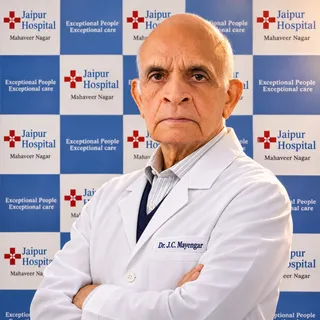
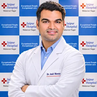
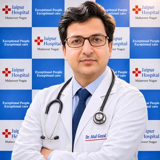
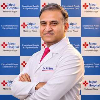
Your orthopaedics doctors
Explore our orthopaedic hospital in Jaipur: every treatment, the doctors and what to expect at your visit.
Frequently asked questions
Questions patients ask about thigh bone (femur) fracture surgery
What is plating and nailing for long bone fracture?
In short
Plating and nailing are surgical methods to fix a broken thigh bone. At Jaipur Hospital, the orthopaedic team uses intramedullary nailing or locking compression plates depending on the fracture pattern. Surgery takes one and a half to three hours under spinal or general anaesthesia. You usually stay in hospital for three to five nights.
In detail
Intramedullary nailing places a metal rod inside the hollow centre of the femur. This shares load with the bone and allows early movement.
Locking compression plates sit on the outer surface and are chosen for fractures near the hip or knee, or when the bone has not healed after previous surgery. Evidence shows reamed nailing lowers implant failure compared with unreamed nailing.
External fixation may be used first for severe open fractures with soft tissue damage. The surgeon selects the method after reviewing X-rays and, if needed, a CT scan done at an outside imaging centre.
A common belief
People often think one method is always better.
What actually happens
In reality, the fracture location and pattern decide which implant gives the most stable fixation.
Your next step at Jaipur Hospital
After surgery, partial weight bearing starts at six to eight weeks. Full recovery takes four to six months with regular physiotherapy. The typical cost range in India is ₹1,20,000 to ₹2,80,000; ask for an estimate based on your implants and stay. Your surgeon will explain the exact plan at the consultation.
Which specialist should I see for this problem?
In short
You need an orthopaedic surgeon who operates on femur fractures. At Jaipur Hospital, the orthopaedic department manages these injuries from emergency admission through surgery and rehabilitation. The team includes surgeons trained in intramedullary nailing and plating techniques.
In detail
Femur shaft fractures require surgical fixation in almost every adult. The orthopaedic surgeon decides between intramedullary nailing, locking compression plate, or temporary external fixation based on fracture location, pattern, and soft tissue condition. The anaesthesia team gives spinal or general anaesthesia.
Physiotherapists start early mobilisation and guide weight-bearing progression. If the fracture is open, the surgeon works with plastic surgery colleagues for soft tissue coverage. All care is coordinated within the hospital.
A common belief
Some patients go to a general physician first.
What actually happens
A femur fracture needs operative fixation within days, so direct referral to orthopaedics saves time and avoids displacement.
Your next step at Jaipur Hospital
Come to the orthopaedic OPD or the 24×7 emergency department at S16A-17, Mahaveer Nagar, Near Gopalpura Flyover, Main Tonk Road, Jaipur. Bring any previous X-rays or reports.
The surgeon will examine you, order imaging if needed, and explain the operation, hospital stay of three to five nights, and recovery timeline.
What treatment options are available for my situation?
In short
The main options are intramedullary nailing, locking plate fixation and temporary external fixation. At Jaipur Hospital the orthopaedic surgeon chooses the method based on fracture location, pattern and soft tissue condition. Intramedullary nailing is standard for most shaft fractures; plates are used near the joints or for non-unions.
In detail
Intramedullary nailing places a rod inside the bone canal, allowing early weight bearing and preserving the fracture haematoma.
Locking compression plates are fixed to the side of the bone with screws and are preferred for proximal or distal fractures where a nail cannot get adequate purchase.
External fixation with pins and an outside frame is a temporary measure for severe open fractures, unstable patients or when definitive surgery must be delayed. Retrograde nailing through the knee is an option for distal femur fractures.
The surgeon discusses the typical operation time of one and a half to three hours, hospital stay of three to five nights and the rehabilitation timeline.
A common belief
Patients sometimes ask for a cast, but it cannot hold a femur fracture straight.
What actually happens
The surgeon selects the implant that gives stable fixation and lets you start moving the knee and hip early.
Your next step at Jaipur Hospital
After imaging review the surgeon explains the recommended fixation, expected recovery of partial weight bearing at six to eight weeks and full recovery in four to six months.
The typical cost range in India is one lakh twenty thousand to two lakh eighty thousand rupees; an estimate is given after the clinical assessment.
What can I safely do at home to manage symptoms of a femur shaft fracture?
In short
While awaiting surgery or during early recovery, keep the injured leg supported on pillows when lying down to reduce swelling. At Jaipur Hospital, the orthopaedic team advises using the prescribed pain medication on schedule, applying ice packs wrapped in a cloth for short periods, and performing ankle pumps and toe movements hourly to promote circulation. Do not put weight on the leg until the surgeon clears you.
In detail
Evidence shows that early mobilisation after stable fixation lowers the risk of blood clots and chest complications. Before surgery, traction may be applied in hospital to maintain length and alignment.
At home, the focus is on protecting the fracture: use crutches or a walker exactly as taught, keep the wound dressing clean and dry, and watch for increasing pain, redness, swelling or fever, which may signal infection. Good nutrition with adequate protein and calories supports bone healing.
A common belief
A common mistake is trying to walk too early or skipping the ankle exercises, which raises the risk of clots and stiffness.
What actually happens
Follow the weight-bearing instructions exactly.
Your next step at Jaipur Hospital
Arrange a ground-floor sleeping area and a bedside commode if stairs are difficult. Ask a family member to help with meals, medication timing and wound checks. Attend all follow-up appointments at Jaipur Hospital for X-ray review and physiotherapy progression.
How will the femur fracture injury be assessed for displacement, instability, or damage to nerves and blood vessels?
In short
At Jaipur Hospital, the orthopaedic team assesses a femur fracture with a physical examination and imaging. The surgeon checks for limb shortening, rotation, swelling, and skin breaks. Pulses, sensation, and movement in the foot are tested to detect nerve or vessel injury. X-rays of the thigh and hip confirm displacement and fracture pattern. If needed, the surgeon may request a CT scan at an outside imaging centre for more detail.
In detail
A femur fracture usually results from severe trauma, and the broken fragments can damage nearby muscles, nerves, and blood vessels. The clinical exam looks for an open fracture where bone pierces the skin, graded by the Gustilo-Anderson system.
Distal pulses and capillary refill show arterial supply; sensation and active movement test the sciatic, femoral, and peroneal nerves. X-rays in two planes reveal displacement, comminution, and shortening.
In complex patterns near the hip or knee, a CT scan helps plan whether intramedullary nailing or plating is appropriate. The surgeon also evaluates the patient's overall condition for other life-threatening injuries before deciding on definitive fixation or temporary external fixation.
Your next step at Jaipur Hospital
Expect a thorough examination and X-rays in the emergency department at Jaipur Hospital. If a CT scan is advised, the team will refer you to a nearby imaging centre and review the images together. Surgery is usually scheduled once you are medically stable.
How will femur fracture treatment affect my walking, movement, or independence?
In short
Femur fracture surgery at Jaipur Hospital aims to restore your ability to walk independently. In the first six to eight weeks you will need a walking frame or crutches for partial weight bearing. Most people regain full weight bearing by four to six months. Some patients report occasional weather-related ache or mild thigh wasting, but long-term studies show the majority return to their previous activities.
In detail
The strong muscles around the femur pull the broken ends out of alignment, so surgery with a nail or plate realigns the bone and holds it while it heals. Early knee and hip movement is encouraged to prevent stiffness.
Evidence shows suprapatellar nailing can reduce anterior knee pain and improve knee scores at six and twenty-four months. However, a small number of patients develop heterotopic ossification around the hip or have rotational malunion that slightly alters gait.
Thigh circumference may be one to two centimetres smaller on the operated side. Full recovery of strength and confidence can take up to a year, especially if you had other injuries.
A common belief
Some patients expect to be back to normal in weeks, but the bone and muscles need months.
What actually happens
A few have a slight limp or knee ache long term, especially if the fracture was complex or they smoke.
Your next step at Jaipur Hospital
Your physiotherapy programme starts in hospital and continues as an outpatient at Jaipur Hospital. You will progress from assisted walking to independent gait as X-rays confirm union. Return to work and driving depend on your job and which leg was operated on.
Which symptoms or changes should prompt me to seek medical advice?
In short
Seek medical help immediately if you have severe thigh pain after a fall or road accident, cannot put weight on the leg, see the leg looking shorter or twisted, or notice swelling and bruising that grows quickly. At Jaipur Hospital, the 24×7 emergency department assesses suspected femur fractures straight away.
In detail
A femur fracture usually follows high-energy trauma such as a vehicle crash or a fall from height. The bone is very strong, so a break means major force was applied.
Symptoms include intense pain, inability to move the leg, obvious deformity, and swelling from bleeding into the thigh. Open fractures show a wound with bone visible. In elderly people with osteoporosis, even a low-energy fall can cause a fracture.
Numbness or tingling in the foot may mean nerve or blood vessel injury. Any of these signs need urgent X-ray and surgical evaluation.
A common belief
Some people wait hoping the pain will settle.
What actually happens
A femur fracture will not heal correctly without surgery, and delay raises the risk of compartment syndrome or blood vessel damage.
Your next step at Jaipur Hospital
Call the ambulance on 7414 001 001 or come to the emergency department at S16A-17, Mahaveer Nagar, Near Gopalpura Flyover, Main Tonk Road, Jaipur. The team will splint the leg, give pain relief, and order X-rays. If surgery is needed, it is usually done within 24 to 48 hours once you are fit for anaesthesia.
What will the doctor ask and check at the first visit?
In short
The orthopaedic surgeon will ask how the injury happened, whether you have other injuries, and about your medical history including medications and allergies. At Jaipur Hospital, the doctor examines the thigh for swelling, deformity, bruising, and checks pulses and sensation in the foot.
In detail
The surgeon needs to know the mechanism — road accident, fall from height, or simple fall — because high-energy trauma often causes other injuries. They will ask about past surgeries, diabetes, osteoporosis, and blood-thinning medicines.
The physical exam looks for leg shortening, rotation, open wounds, and compartment tightness. Distal pulses and nerve function are documented. X-rays of the femur, hip and knee are ordered immediately.
If the fracture extends near a joint, a CT scan at an outside imaging centre may be requested for surgical planning.
A common belief
Patients sometimes forget to mention blood thinners or diabetes.
What actually happens
These affect anaesthesia and infection risk, so the surgeon must know before planning surgery.
Your next step at Jaipur Hospital
After the assessment, the surgeon explains the fixation method, expected hospital stay of three to five nights, and the recovery phases. You will be told about anaesthesia options and the need for physiotherapy. Consent is taken once you understand the plan.
Can a femur shaft fracture improve without a procedure or operation?
In short
A femur shaft fracture usually needs surgery to heal correctly. At Jaipur Hospital, the orthopaedic team advises that non-operative care is rarely suitable because the thigh bone bears heavy loads and strong muscle pull. Without fixation, the bone ends often shorten, rotate or angulate, leading to long-term limp and joint wear.
In detail
Evidence shows that intramedullary nailing is the standard treatment for most adult femoral shaft fractures because it restores alignment and allows early movement. In studies, non-operative management such as traction or casting results in higher rates of malunion, shortening and knee stiffness.
Surgery also reduces the time spent bed-bound, which lowers the risk of chest infection, blood clots and pressure sores. The surgeon will consider your fracture pattern, bone quality and general health before recommending the exact implant.
A common belief
People sometimes think traction or a long plaster will work, but studies show that leads to shortening, twisting and a stiff knee.
What actually happens
Surgery with a nail or plate gives reliable alignment and lets you start moving sooner.
Your next step at Jaipur Hospital
If you have a suspected femur fracture, attend the emergency department at Jaipur Hospital for X-ray assessment and pain relief. The team will explain whether surgery is advised and discuss the typical hospital stay of three to five nights.
Are there foods, activities, or habits I should change for a femur shaft fracture?
In short
Eat a balanced diet rich in protein, calcium and vitamin D to support bone healing. At Jaipur Hospital, the orthopaedic team recommends stopping smoking completely, as nicotine delays bone union and raises infection risk. Limit alcohol, avoid putting weight on the operated leg until cleared, and follow the physiotherapy exercise programme daily. Do not drive while you are on strong pain medication or cannot bear weight safely.
In detail
Evidence links smoking to higher nonunion rates and longer healing times. Protein intake of 1.2 to 1.5 grams per kilogram of body weight per day is often advised during fracture healing.
Calcium and vitamin D supplementation may be prescribed if dietary intake is low or blood levels are insufficient. High-impact activities, contact sports and heavy lifting are restricted until radiographic union is confirmed, typically around four to six months. The surgeon will guide the gradual return to work based on your job demands.
A common belief
Many people underestimate how much smoking and poor nutrition delay healing.
What actually happens
Studies show smokers have significantly higher nonunion rates. The daily physio routine is not optional; it prevents stiffness and muscle loss.
Your next step at Jaipur Hospital
Ask the dietician at Jaipur Hospital for a meal plan if you have dietary restrictions. Set reminders for physiotherapy sessions and medication. Keep a simple log of pain scores and walking distance to discuss at follow-up.
FAQ library
Questions and answers about thigh bone (femur) fracture surgery
When you contact us
Get a clear plan and cost estimate
Contact Jaipur Hospital for an OPD consultation where your scans are reviewed, options explained and a cost estimate given before you decide.
- A consultation with an orthopaedic surgeon in the OPD
- Your scans and reports reviewed, with tests done in-house if you need them (laboratory, X-ray and imaging)
- Your options explained in plain words, including whether you need surgery at all
- A cost estimate before you decide
- Help with cashless treatment under MAA Yojana, various government health care schemes and empanelled insurers and TPAs
- Follow-up visits after your treatment
Overview
Thigh Bone (Femur) Fracture Surgery: what to know
What a femur fracture is and how it happens
- Thigh bone
- Longest and strongest bone in the body
- High-energy injury
- Road crash or fall from height in adults
- Low-energy fall
- Standing-height fall in older people with thin bones
- Open or closed
- Skin intact or bone pierces the skin
The femur is your thigh bone. It is the longest and strongest bone in your body. A femur fracture is the medical term for a broken thigh bone. Because the bone is so strong, it usually takes a severe force to break it.
In most adults the break follows a high-energy injury such as a road traffic crash or a fall from height. In older people, especially women with thinner bones, a simple fall from standing height can be enough to cause the fracture. The break may be closed, where the skin stays intact, or open, where the bone pierces the skin. The fragments may stay in line or be displaced, leaving a gap between the broken ends.
Symptoms of a thigh bone fracture and when to seek urgent care
- Severe thigh pain Immediate and intense
- Cannot bear weight Unable to put weight on the injured leg
- Leg looks deformed Shorter, twisted or bent at an odd angle
- Open wound with bleeding Bone has broken through the skin
- Numb or cold foot Possible nerve or blood vessel injury
A femur fracture causes immediate, severe pain in the thigh. You will not be able to put weight on the injured leg. The leg may look shorter, twisted or bent at an odd angle. Swelling and bruising appear quickly. If the bone has broken through the skin you will see an open wound with bleeding.
Numbness or a cold foot can mean nerve or blood vessel injury. Any of these signs need emergency care the same day. Call an ambulance or go straight to the nearest emergency department.
Causes and people at higher risk of a femur fracture
- High-energy trauma
- Car crashes, falls from height, sports injuries
- Osteoporosis
- Fragile bone after menopause
- Other bone-weakening factors
- Osteopenia, long-term steroids, bone-weakening conditions
- Open fractures
- Higher chance of infection and complications
High-energy trauma such as car or motorcycle crashes, falls from height, and sports injuries cause most femur shaft fractures in younger adults. In older adults, especially women after menopause, osteoporosis makes the bone fragile so a low-energy fall from standing height can break the femur.
People with osteopenia, long-term steroid use, or conditions that weaken bone are also at higher risk. Open fractures are less common but carry a higher chance of infection and other complications.
How a femur fracture is diagnosed: examination and imaging
- Examination Check deformity, swelling, wounds, pulses, sensation
- X-rays first Thigh and knee X-rays usually confirm the fracture
- CT or MRI if needed Done at an outside imaging centre, reviewed by your surgeon
- Fracture description Open or closed, displaced or not, location on shaft
In the emergency department a doctor examines your leg for deformity, swelling, wounds, pulses and sensation. X-rays of the thigh and knee are the first imaging test and usually confirm the fracture. A CT scan or MRI may be advised to see the fracture pattern more clearly, especially near the hip or knee joints, or to plan surgery.
These scans are done at an imaging centre outside the hospital and the reports are reviewed by your surgeon. The fracture is described as open or closed, displaced or non-displaced, and by its location along the shaft.
Non-surgical treatment and when surgery is advised for a femur fracture
Almost all femur shaft fractures in adults need surgery to heal correctly. Without fixation the bone fragments usually shorten, rotate or angulate, leading to a limp, leg-length difference and long-term knee or hip problems. Traction with weights and splints may be used for a short time before surgery if you have other life-threatening injuries or are not fit for anaesthesia.
External fixation with pins and an outside frame is sometimes applied as a temporary measure in severe open fractures or when definitive surgery must wait. Definitive fixation is either an intramedullary nail placed inside the bone canal or a locking plate fixed to the side of the bone. Your surgeon will advise which method suits your fracture pattern, age, bone quality and any other injuries.
Preparing for Thigh Bone (Femur) Fracture Surgery
Before the operation your surgical team will review your general health. You will have blood tests, an ECG and a chest X-ray to check that you are fit for anaesthesia. If you take blood-thinning medicines, diabetes tablets or insulin, or any regular medication, tell your surgeon so the dose can be adjusted or stopped for a short time.
You must not eat or drink for at least six hours before the planned surgery time. Bring your regular medicines, a list of allergies, your insurance or scheme cards (MAA Yojana, various government health care schemes or private TPA details) and comfortable clothes for discharge. An attendant should stay with you during the admission. The emergency department and ambulance service run 24 hours a day if you arrive after a fresh injury.
How Thigh Bone (Femur) Fracture Surgery Is Done
- Anaesthesia Spinal or general anaesthesia
- Fixation choice Nail, plate or external fixator based on fracture pattern
- Intramedullary nailing Metal rod down the centre, locked with screws at both ends
- Locking plate Used near hip or knee, or after failed previous surgery
- External fixation Temporary for severe open fractures or unfit patients
- Retrograde nailing Rod inserted from the knee end for fractures near the knee
The operation is performed under spinal or general anaesthesia and usually takes one and a half to three hours. Your surgeon will choose the fixation method that fits your fracture pattern.
Intramedullary nailing is the standard treatment for most femoral shaft fractures in adults; a metal rod is passed down the centre of the bone and locked with screws at both ends. A locking compression plate may be used for fractures near the hip or knee, or when the bone has not healed after a previous operation.
External fixation with pins and an outside frame is sometimes applied as a temporary measure for severe open fractures with extensive soft-tissue damage, or when your condition does not allow a longer operation. Retrograde nailing, where the rod is inserted from the knee end, is an option for fractures just above the knee joint.
On the day of surgery you are taken to the modular operation theatre, the skin is cleaned, and the chosen implants are placed using X-ray guidance. The wound is closed in layers and a sterile dressing is applied. You then go to the recovery area and, once stable, to the ward.
Risks and Possible Complications of Femur Fracture Surgery
- Over 30% with systemic antibiotics alone; below 9% with local antibiotics added Deep infection (open fractures)
- About 6% Non-union
- About 2% Delayed union
- Reamed nailing showed fewer failures than unreamed Implant failure
All operations carry some risk. Infection can occur at the wound or around the implant; in studies of severe open fractures the deep infection rate was over 30 percent with systemic antibiotics alone, but fell below 9 percent when local antibiotics were added at the implant site.
Implant failure such as broken screws or nail breakage has been reported; reamed nailing showed fewer implant failures than unreamed nailing in large trials. Non-union, where the bone does not heal, occurred in about 6 percent of patients in some series, and delayed union in about 2 percent.
Malunion, where the bone heals with an angle or shortening, was seen in a small number of cases, including rotational malunion and a few centimetres of shortening. Nerve injury such as pudendal nerve palsy has been described and usually recovers over time. Heterotopic ossification (extra bone forming around the hip) was noted in a few patients after nailing.
The injuries that cause the fracture can also damage muscles, blood vessels and other bones, and the broken fragments may injure surrounding tissue when they move. Your surgical team will discuss the risks that apply to your fracture type and general health.
Recovery and Aftercare After Femur Fracture Surgery
- Hospital stay Three to five nights
- Partial weight bearing Starts at six to eight weeks with X-ray signs of healing
- Full recovery Four to six months, up to a year for all activities
- Physiotherapy Several months to rebuild strength and movement
You will stay in hospital for three to five nights. Pain is managed with regular medication. A physiotherapist will teach you how to move safely with a walking frame or crutches. Partial weight bearing usually starts at six to eight weeks, guided by X-ray signs of healing.
Full recovery takes four to six months, and it can take up to a year to regain all your previous activities. You will need several months of physiotherapy to rebuild strength and joint movement. Do not start sports, heavy work or intense exercise until your doctor confirms it is safe.
Keep the wound clean and dry; stitches or clips are removed at the follow-up visit. Contact the hospital if you develop increasing pain, swelling, redness, discharge, fever, sudden shortness of breath, or if the leg becomes pale, cold or numb. The 24-hour emergency department is available for urgent concerns.
Results of Femur Fracture Surgery in Studies
In a large multicentre trial involving over 1,300 participants, there was no statistically significant difference in major re-operation rates between reamed and unreamed nailing. Non-union, deep infection and implant failure rates were also similar between the two methods. A subgroup analysis suggested reamed nailing may reduce major re-operations related to non-union in closed fractures.
Expandable nails showed a lower re-operation rate than standard interlocking nails in one trial. The suprapatellar nailing approach was associated with less anterior knee pain, better knee scores and shorter X-ray time compared with the infrapatellar approach, without increasing operation time, blood loss or hospital stay.
Overall healing rates of 93 to 94 percent have been reported in series with mean healing times around 19 weeks. Most patients regain near-normal hip and knee motion, although a small number report weather-related aching or discomfort at the implant site in the longer term.
Cost and Insurance for Femur Fracture Surgery in Jaipur
The typical price range for femur fracture fixation at Jaipur Hospital is ₹1,20,000 to ₹2,80,000. The final cost depends on the type of implant (nail or plate), the complexity of the fracture, the length of stay, and whether additional procedures such as wound debridement or bone grafting are needed.
The hospital accepts MAA Yojana, various government health care schemes and a panel of private insurers and TPAs; bring your scheme or policy documents at admission. The in-house pharmacy and diagnostic laboratory are available round the clock. For a detailed estimate based on your specific case, speak to the admissions desk.