Orthopaedics · Jaipur Hospital
High Tibial Osteotomy in Jaipur
Knee pain from early arthritis? Jaipur Hospital offers assessment and a clear plan.
- Serving Jaipur since 1991 (35 years)
- NABH accredited
- 24×7 emergency & ICU
- Cashless: MAA Yojana + 32 insurers
- Operation time
- 1.5 to 2.5 hours
- Anaesthesia
- Spinal or general anaesthesia
- Recovery
- Full weight bearing in 8 to 12 weeks, return to sports in 4 to 6 months
- Price
- ₹1,50,000 to ₹3,00,000typical range in India; ask us for an estimate
Knee pain limits daily life
Pain on the inner knee that worsens with walking or stairs
Early arthritis wears the inner side of the knee. The leg bows inward, shifting weight onto the damaged area. Pain, stiffness and swelling make walking, squatting or standing for long periods difficult.
-
Pain with activity
Aching or sharp pain on the inner knee when walking, climbing stairs or standing.
-
Morning stiffness
Knee feels tight and hard to move after sitting or waking up.
-
Leg bowing inward
The knee angles inward, putting more pressure on the worn cartilage.
-
Swelling after use
The knee puffs up after a long day or exercise.
-
Limits work and home tasks
Difficulty squatting, kneeling or walking distances needed for daily chores.
Knee osteoarthritis has biomechanical and inflammatory origins and is worsened by excess weight.
Why Jaipur Hospital
You worry that knee pain will force you to give up work, household duties or the activities that keep you independent.
Your care plan
Your first steps with us
- 1 Call or visit OPD for surgeon consultation
- 2 Review scans and plan the corrective surgery
- 3 Admit for osteotomy with spinal or general anaesthesia
- 4 Begin guided physiotherapy and gradual weight bearing
How the operation is done
The surgeon cuts the upper tibia and changes its angle to shift weight from the worn inner side to the healthier outer side. The bone is fixed with a plate and screws. The procedure takes 1.5 to 2.5 hours with a 2 to 3 night stay.
-
Medial Opening Wedge Osteotomy
A wedge is opened on the inner side of the tibia and held with a plate and screws. No bone is removed.
-
Lateral Closing Wedge Osteotomy
A wedge of bone is removed from the outer side and the gap closed, fixed with a plate.
-
Combined with Microfracture
Small holes are made in the damaged cartilage area during the same surgery to stimulate repair tissue.
Why patients trust us
- 1991 Serving Jaipur for 35 years
- NABH Accredited hospital
- 4 Orthopaedics consultants
- 24×7 Emergency, ICU and ambulance
- 33 Cashless schemes and insurers
What research shows about this treatment
-
About 8 in 10 return to work by one year
In study groups, more than 80% of patients went back to work, mainly within 12 months after surgery.
-
About 8 in 10 return to sport at same or higher level
Approximately 80% of patients in studies returned to sport at a level equal to or better than before surgery.
-
Non-union occurs in about 4 in 100 cases
Reported non-union rates range from 0.7% to 4.4% across different fixation methods in published series.
If it is left untreated
Without realignment, the worn inner cartilage continues to thin. Pain and stiffness increase, walking distance shortens, and the leg bows further. This often leads to full knee replacement at a younger age.
Life after treatment
Most people walk without support by three months and resume light sport by six months. The knee feels more stable and daily tasks become easier.
-
Walk without aid
Most people stop using crutches or a stick around 8 to 12 weeks after surgery.
-
Return to light sport
Cycling, swimming or badminton often resume by 4 to 6 months with guided rehab.
-
Easier daily tasks
Squatting, kneeling and climbing stairs usually improve as strength returns.
-
Delay joint replacement
The realignment buys years before a total knee replacement may be needed.
Meet the team
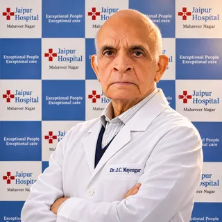
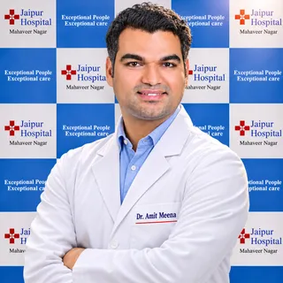
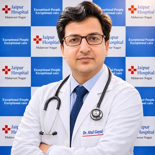
Your orthopaedics doctors
Explore our orthopaedic hospital in Jaipur: every treatment, the doctors and what to expect at your visit.
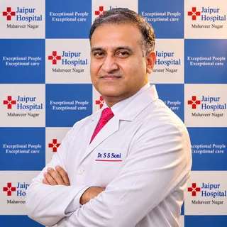
Frequently asked questions
Questions patients ask about high tibial osteotomy
What does high tibial osteotomy involve?
In short
High tibial osteotomy is a bone realignment surgery for early knee arthritis affecting the inner side of your joint. At Jaipur Hospital, our surgical team realigns your shinbone to shift body weight onto the healthier side. The procedure usually takes 1.5 to 2.5 hours under spinal or general anaesthesia.
In detail
Surgical techniques include opening a bone wedge on the inner side or removing a wedge from the outer side, followed by fixation with a metal plate and screws. The operation preserves your natural joint and aims to relieve load imbalance.
In studies, gaps under 10 millimetres filled with new bone over 6 to 18 months without requiring a bone graft. When focal cartilage damage is also present, your surgeon may combine the realignment with microfracture during the same sitting.
In published research, overall long-term survivorship ranges from 40% to 85%, delaying or preventing the need for a total knee replacement.
A common belief
Many people assume knee arthritis always means replacing the joint with artificial parts.
What actually happens
High tibial osteotomy preserves your natural knee, keeping your own bone and ligaments intact while relieving pressure on worn cartilage.
Your next step at Jaipur Hospital
You typically stay in hospital for 2 to 3 nights with your attendant. Full weight bearing usually takes 8 to 12 weeks, while returning to sports takes 4 to 6 months. In studies, over 80% of patients returned to work within a year. You can book an orthopaedic consultation at Jaipur Hospital to discuss whether bone realignment suits your knee.
Which tests and scans do I need beforehand for high tibial osteotomy?
In short
You need standing X-rays of your legs and routine pre-operative health checks before high tibial osteotomy. At Jaipur Hospital, our team performs your blood tests, urine tests, and an ECG. These checks confirm your fitness for anaesthesia and measure the precise angle needed for bone realignment.
In detail
Long-leg alignment X-rays show the mechanical axis from your hip to your ankle, helping your surgeon plan the exact bone wedge correction. Basic blood counts, kidney function tests, and blood sugar tests assess your overall healing capacity and infection risk.
While our in-house radiology provides standard X-rays, Jaipur Hospital does not have a CT or MRI scanner. If your surgeon advises an MRI to inspect soft tissues or cruciate ligaments, you will complete it at an outside imaging centre and bring the films. Your surgeon reviews these scans alongside standard pre-anaesthetic health assessments before surgery.
A common belief
People often think a routine standard knee X-ray is enough.
What actually happens
Planning a bone realignment requires full-length leg X-rays to calculate your mechanical axis, and sometimes an outside MRI to check your cartilage and ligaments.
Your next step at Jaipur Hospital
Pre-operative testing is typically scheduled a few days before your planned admission. Your surgical team will guide you through each assessment and outline expected timelines. Typical hospitalisation lasts 2 to 3 nights, and recovery to full weight bearing takes 8 to 12 weeks. You can visit Jaipur Hospital to complete your pre-operative orthopaedic workup.
How painful is high tibial osteotomy and the early recovery?
In short
High tibial osteotomy involves surgical bone cutting, so you will experience surgical pain and swelling during early recovery. At Jaipur Hospital, your surgical team uses anaesthesia during the operation and prescribes pain medicines to help you stay comfortable while your leg begins healing over the first few days.
In detail
The operation is carried out under spinal or general anaesthesia, meaning you feel no pain while the bone realignment takes place. When the anaesthesia wears off, surgical soreness is normal as tissues heal around the cut bone and fixation plate.
In studies, high tibial osteotomy entails a relatively high overall minor complication rate, but serious complications remain low. In studies, patients who maintained successful joint survival reported a mean Knee injury and Osteoarthritis Outcome Score Pain score of 91, alongside high functional daily living scores. Pain levels steadily lessen as the bone consolidation advances over the initial weeks.
A common belief
Some people fear unbearable long-term agony, while others expect zero discomfort.
What actually happens
The truth is that early bone healing causes temporary ache, but in published studies, long-term pain scores reach an average of 91 out of 100 once the joint heals.
Your next step at Jaipur Hospital
You will typically spend 2 to 3 nights in the hospital, where our team monitors your pain and helps you mobilise safely. After discharge, take your prescribed pain tablets from the chemist exactly as advised. Keep your leg elevated to reduce throbbing and swelling. To discuss pain management and surgical planning for knee realignment, visit Jaipur Hospital for a consultation.
How long does recovery from high tibial osteotomy usually take?
In short
Recovery from high tibial osteotomy usually requires several months for bone healing, walking rehabilitation, and muscle strengthening. At Jaipur Hospital, patients typically achieve full weight bearing in 8 to 12 weeks and return to recreational sports activities within 4 to 6 months under physiotherapist guidance.
In detail
The operation takes 1.5 to 2.5 hours and typically involves a hospital stay of 2 to 3 nights. In the initial weeks, you use crutches to protect the healing bone cut and internal fixation plate.
Medical studies show that gaps under 10 millimetres in opening wedge procedures show progressive ossification filling at least 75% within 6 to 18 months.
In published recovery studies, more than 80% of patients returned to work, mainly by 1 year postoperatively, and approximately 80% returned to sport at a level equal to or greater than before surgery.
- Medical studies show that gaps under 10 millimetres in opening wedge procedures show progressive ossification filling at least 75% within 6 to 18 months.
A common belief
Some expect to walk freely within days, while others fear a year of bed rest.
What actually happens
In reality, crutches protect the cut bone initially, and studies show over 80% of patients return to work within one year.
Your next step at Jaipur Hospital
You will follow a graded physiotherapy programme to restore knee bending, quadriceps strength, and steady walking. Desk workers often resume work earlier than manual labourers. Keep attending your planned follow-up reviews to track bone union on X-rays.
To understand your personal recovery roadmap and schedule your assessment, book an appointment with our specialist surgical team at Jaipur Hospital.
How will foot shape, joint alignment, strength, and walking be assessed before high tibial osteotomy?
In short
Our surgical team evaluates your walking pattern, limb alignment, and joint stability during an OPD consultation at Jaipur Hospital. Clinical assessment checks whether varus malalignment shifts excess body weight onto the inner side of your knee, while in-house standing X-rays confirm your bone structure and deformity angles.
In detail
Assessment checks if bone realignment surgery suits your knee arthritis. In studies, high tibial osteotomy corrects varus malalignment in medial compartment knee osteoarthritis, shifting load off the damaged inner compartment.
The specialist team measures your joint line obliquity and checks for ligament laxity, such as chronic posterolateral corner deficiency, which can cause abnormal thrust during walking. Correct patient selection is essential for good long-term results.
Surgical candidates are typically under 55 years of age, active, and not obese. If complex scans like an MRI or CT are advised to check cartilage or soft tissues, they are done at an outside imaging centre.
A common belief
Many people assume knee pain requires immediate joint replacement.
What actually happens
However, careful alignment checks often show that early arthritis affects only the inner side, which can be protected by realigning the bone.
Your next step at Jaipur Hospital
Your evaluation begins in the orthopaedic OPD with a physical check-up and long-leg alignment radiographs. If previous medical reports or X-rays are available, bring them along for your initial review.
Following this thorough evaluation, your surgeon discusses whether realignment surgery or non-surgical care suits your knee. You can book an OPD assessment with our specialist surgical team at Jaipur Hospital to check your joint alignment.
How will high tibial osteotomy affect my walking, movement, or independence?
In short
High tibial osteotomy straightens bowed limb alignment, relieving inner joint pain and improving long-term walking stability at Jaipur Hospital. While your independence is briefly reduced during the initial recovery weeks while using walking aids, everyday movement and physical function improve substantially once your bone heals completely.
In detail
By realigning the tibia, the procedure shifts mechanical weight toward the healthier outer side of the knee.
In clinical trials, patients who underwent realignment reported significant reductions in daily pain and marked gains in joint function, achieving average activities of daily living scores of 97 and pain scores of 91 on validated outcome scales.
Correcting underlying varus alignment also stabilises the knee during gait, especially in patients with associated ligament strain or abnormal joint line tilt.
Although temporary assistance from an attendant is needed during your 2 to 3 night hospital stay and early home recovery, long-term physical independence is restored.
Your next step at Jaipur Hospital
During your first few weeks at home, simple preparations help maintain comfort and safety while your mobility is restricted. Physiotherapy exercises taught in the ward will help you regain knee flexion and quadriceps strength. As bone consolidation progresses over 8 to 12 weeks, independent walking resumes without external support.
Why might my doctor recommend high tibial osteotomy?
In short
Your doctor may recommend high tibial osteotomy to treat early arthritis confined to the inner side of your knee joint. At Jaipur Hospital, our surgical team evaluates your alignment to relieve joint strain. Realigning the shinbone shifts your weight to healthy cartilage, helping preserve your natural joint.
In detail
Knee osteoarthritis causes chronic pain and stiffness, often worsened by extra body weight and varus alignment, commonly known as bowlegs. Straightening the bone corrects the mechanical axis and unloads the worn medial compartment.
In clinical studies, this procedure has historically served as a definitive treatment strategy for medial arthritis, normalising joint obliquity and restoring stability. Research reports 10-year joint survival rates ranging between 51% and 93.2%.
By realigning your leg mechanically, the surgery reduces daily pain while allowing active individuals to maintain higher physical function without replacing the knee joint with artificial metal components.
A common belief
A joint replacement removes bone and inserts metal implants, which can wear out over decades in younger people.
What actually happens
High tibial osteotomy protects your natural joint, letting you stay active and postpone artificial joint surgery.
Your next step at Jaipur Hospital
The surgery typically requires 2 to 3 nights in hospital and 8 to 12 weeks before full weight bearing. Studies show about 80% of patients return to sports at or above previous levels. Typical surgical costs in India range from ₹1,50,000 to ₹3,00,000.
How should I prepare in the days before high tibial osteotomy?
In short
Preparing for high tibial osteotomy involves completing medical clearances, arranging home recovery support, and maintaining basic joint movement. At Jaipur Hospital, our specialist team provides clear pre-operative instructions to help you organise your admission and plan for post-surgery mobility safely.
In detail
Physical preparation focuses on muscle strength and general health. Clinical evidence demonstrates that dietary adjustments combined with light exercise significantly reduce knee joint compressive forces and lower systemic inflammatory markers such as interleukin-6.
Strengthening your upper body and quadriceps helps you manage crutches or a walker during the early non-weight-bearing phase.
You should prepare your home by clearing walkways, setting up a ground-floor resting area, and arranging for a family attendant to stay with you during your hospital stay of 2 to 3 nights.
A common belief
Preparation is not just about showing up on the day.
What actually happens
Setting up a single-floor living space and practicing with walking aids beforehand makes the 8 to 12 week recovery far smoother.
Your next step at Jaipur Hospital
Stop smoking, follow bathing instructions, and purchase prescribed assistive devices ahead of time. You should also finalise your insurance paperwork through our hospital desk. The procedure generally costs ₹1,50,000 to ₹3,00,000 in India.
What are the common side effects and the less common serious complications of high tibial osteotomy?
In short
Common side effects of high tibial osteotomy include temporary knee swelling, bruising, and minor surgical wound soreness. At Jaipur Hospital, our surgical team explains all potential outcomes clearly. While minor problems occur relatively often, serious complications are infrequent when the operation and recovery follow standard medical protocols.
In detail
In medical studies, minor complications are relatively frequent, including superficial wound infections treated in the community with oral antibiotics in about 9.5% of cases. Infections requiring hospital readmission occur in roughly 3.5% of patients.
Bone healing difficulties or non-union range between 0.7% and 4.4% in published reports. Less common complications include blood clots or nerve injury; specifically, common peroneal nerve injury has been described in 2% to 16% of lateral closing wedge procedures, causing foot weakness.
Internal plate fixation maintains infection rates below or equal to 4%. Our surgical team monitors your incision and alignment meticulously to minimise these surgical risks.
- In medical studies, minor complications are relatively frequent, including superficial wound infections treated in the community with oral antibiotics in about 9.5% of cases.
- Infections requiring hospital readmission occur in roughly 3.5% of patients.
A common belief
People often worry about major bone failure.
What actually happens
However, non-union happens in only 0.7% to 4.4% of cases in studies, and most minor infections clear up quickly with simple antibiotic tablets from the chemist.
Your next step at Jaipur Hospital
Mild swelling and leg stiffness are typical and improve over several weeks with guided physiotherapy. Report any high fever, worsening calf pain, or wound drainage immediately to our emergency department. Regular follow-up appointments allow prompt evaluation of bone healing. To review the benefits and potential risks of knee realignment, book a consultation at Jaipur Hospital.
Which symptoms are expected after high tibial osteotomy, and which need urgent medical attention?
In short
Mild swelling, skin bruising, and moderate incision tenderness are normal expected symptoms after high tibial osteotomy. At Jaipur Hospital, our surgical team advises you to seek urgent medical care if you develop high fever, increasing calf pain, severe redness, or sudden leg numbness.
In detail
Following bone realignment, local fluid retention and bruising around the knee or lower leg are standard bodily reactions. However, serious complications can happen.
In medical research, deep surgical site infections requiring hospital readmission occurred in 3.5% of cases, while 9.5% developed minor superficial wound infections needing oral antibiotics.
In addition, nerve injury such as common peroneal nerve palsy has been documented in 2% to 16% of lateral closing wedge procedures.
Sudden difficulty lifting your foot, foul-smelling wound discharge, chest tightness, or calf tenderness require rapid evaluation to rule out nerve damage, deep infection, or blood clots.
Your next step at Jaipur Hospital
Your attendant will receive instructions on warning signs before discharge, typically after 2 to 3 nights in hospital. If you notice persistent fever or worsening wound drainage, visit our 24x7 emergency department immediately. Routine check-ups occur in our OPD. For clear advice on symptom monitoring after high tibial osteotomy, book a consultation at Jaipur Hospital.
FAQ library
Questions and answers about high tibial osteotomy
When you contact us
Get a clear plan for your knee
Contact Jaipur Hospital for an OPD consultation, scan review, cost estimate and help with cashless schemes.
- A consultation with an orthopaedic surgeon in the OPD
- Your scans and reports reviewed, with tests done in-house if you need them (laboratory, X-ray and imaging)
- Your options explained in plain words, including whether you need surgery at all
- A cost estimate before you decide
- Help with cashless treatment under MAA Yojana, various government health care schemes and empanelled insurers and TPAs
- Follow-up visits after your treatment
Overview
High Tibial Osteotomy: what to know
What is medial compartment knee arthritis and how it happens
- Worn inner cartilage
- Thigh and shin bones rub together
- Bowed leg shape
- Varus alignment shifts weight inward
- Faster wear inside
- Inner cartilage wears before outer side
- Joint space narrows
- Bone spurs may form over time
Medial compartment knee arthritis means the smooth cartilage on the inner side of the knee joint has worn away. The thigh bone and shin bone then rub against each other when you walk or stand. This causes pain, stiffness and swelling. The problem often starts because the leg is slightly bowed, a shape called varus alignment.
That shape pushes more body weight through the inner side of the knee, so the cartilage there wears faster than the outer side. Over time the joint space narrows and bone spurs may form.
High tibial osteotomy (HTO) is a joint-preserving operation that realigns the upper shin bone. By changing the angle of the bone, weight is shifted from the damaged inner side to the healthier outer side of the knee. This can relieve pain and delay or avoid the need for a knee replacement.
Symptoms of medial knee arthritis and warning signs that need urgent care the same day
- Sudden severe pain Cannot bear weight on the leg
- Hot, red, swollen knee with fever Can indicate joint infection
- Numbness or weakness in the foot May point to nerve injury
Typical symptoms include pain on the inner side of the knee, especially when you put weight on the leg. The knee may feel stiff after sitting for a while and may swell after activity. You might notice a bow-legged appearance that gets more obvious over time. Walking distance often becomes limited and climbing stairs can be difficult.
Some signs need medical attention on the same day. Sudden severe pain with inability to bear weight may mean a fracture or a sudden tear. A hot, red, very swollen knee with fever can indicate infection in the joint. Numbness or weakness in the foot may point to nerve injury. If any of these appear, go to the emergency department at Jaipur Hospital or the nearest hospital without delay.
Causes and who is at higher risk for medial knee arthritis
- Not obese
- BMI 25 to 27.5 linked with suitable results
- Inner compartment only
- Arthritis limited to medial side
- Intact ligaments and good motion
- Correctable deformity also needed
The main cause is abnormal load on the inner knee joint. A varus (bow-legged) alignment shifts weight medially. Previous injury to the meniscus or ligaments on that side can change how forces travel through the knee. Obesity adds extra load and also increases inflammation in the joint.
People most likely to benefit from high tibial osteotomy are those under 55 years of age, who are not obese, and whose arthritis is limited to the inner compartment without severe disability. Studies show that patients with a body mass index between 25 and 27.5 had the most suitable results.
Age around 45 was linked with better long-term survival of the osteotomy. Good candidates also have intact ligaments, reasonable knee movement and a correctable deformity.
How medial knee arthritis is diagnosed: examination and tests
Your surgeon will ask about your pain pattern, walking distance and any past injuries. The examination checks leg alignment, knee range of motion, ligament stability and where the joint line is tender. Standing X-rays of both legs are the main test. They show the joint space narrowing on the inner side and the overall mechanical axis of the leg. Special long-leg alignment views help measure the exact correction needed for surgery.
Jaipur Hospital has an in-house X-ray facility. CT and MRI scans are not available at the hospital; if your surgeon advises them, they are done at an imaging centre outside and the images are brought back for review. Blood tests may be ordered to rule out inflammatory arthritis or infection before any operation.
Treatment options without surgery and when high tibial osteotomy is advised
Non-surgical treatment is tried first. Weight loss reduces load on the knee; in one trial a combined diet and exercise programme achieved about 10 kg mean weight loss and lowered inflammatory markers and knee compressive forces. Physiotherapy strengthens the thigh muscles and improves joint control.
A knee brace or a lateral wedge insole can unload the inner compartment for some people. Pain medicines such as paracetamol or NSAIDs help manage symptoms but do not change the disease.
High tibial osteotomy is advised when the arthritis is mainly in the inner compartment, the leg is in varus, the ligaments are intact, knee movement is reasonable, and non-surgical measures no longer control pain or allow daily activity. It is a joint-preserving choice for patients who are too young or too active for a knee replacement.
The two common techniques are medial opening wedge osteotomy and lateral closing wedge osteotomy; your surgeon will explain which suits your anatomy. If a focal cartilage defect is found, microfracture can be done at the same time.
Preparing for high tibial osteotomy
Do
- Recent knee X-rays and MRI reports
- List of current medicines
- Insurance cards
- Comfortable loose clothing
- Attendant to stay during admission
Before high tibial osteotomy your surgeon will review your medical history and current medicines. Blood tests, an ECG and a chest X-ray are usually done to check fitness for anaesthesia. If you take blood thinners, diabetes tablets or insulin, your doctor will tell you when to stop or adjust them.
You will be asked to fast for at least six hours before the operation. Bring any recent knee X-rays, MRI reports, a list of medicines, your insurance cards and comfortable loose clothing. An attendant should plan to stay with you during the hospital stay.
How high tibial osteotomy is done
High tibial osteotomy is performed under spinal or general anaesthesia and takes about one and a half to two and a half hours. The surgeon makes a cut below the knee and reshapes the upper tibia to shift weight away from the worn inner compartment. Three main techniques are available.
In a medial opening wedge osteotomy the bone is opened on the inner side and held with a plate and screws; gaps smaller than 10 mm are often left to heal without graft. In a lateral closing wedge osteotomy a wedge of bone is removed from the outer side and the gap is closed with a plate.
If a focal cartilage defect is found, microfracture can be done at the same time to encourage repair tissue. Your surgeon will advise which technique suits your alignment, bone quality and activity level. The hospital uses standard instruments and alignment guides; robotic or computer-navigated systems are not used here.
A CT or MRI scan may be advised before surgery; these are done at an imaging centre outside the hospital and the surgeon reviews the images.
Risks and possible complications of high tibial osteotomy
- 31 percent Overall complication rate
- 9.5 percent Minor wound infections
- 3.5 percent Infections needing admission
- 0.7 to 4.4 percent Non-union rate
High tibial osteotomy has a low serious complication rate but a relatively high overall minor complication rate. In one large review the overall complication rate was 31 percent. Minor wound infections needing oral antibiotics occurred in about 9.5 percent of patients, while infections requiring hospital admission were seen in 3.5 percent.
Non-union, where the bone does not heal, has been reported between 0.7 percent and 4.4 percent; in one series the overall non-union rate was 4.3 percent and all cases went on to heal after revision. Infection rates with internal fixation are generally 4 percent or less.
Common peroneal nerve palsy after lateral closing wedge osteotomy has been described in 2 to 16 percent of cases. Patella baja, a lowering of the kneecap due to tendon contracture, was reported in 7.6 to 8.8 percent of patients when long-term cast immobilisation was used. Your surgical team will discuss the risks that apply to your technique and health.
Recovery and aftercare after high tibial osteotomy
- Hospital stay Two to three nights
- Hinged knee brace Worn for first six weeks
- Partial weight bearing Starts soon after surgery with crutches
- Full weight bearing Over eight to twelve weeks as bone heals
- Desk work return Often by six to eight weeks
- Heavy manual work May take three to four months
You will stay in hospital for two to three nights. Pain is managed with regular medication and ice packs. A hinged knee brace is usually worn for the first six weeks. You will start partial weight bearing with crutches soon after surgery and progress to full weight bearing over eight to twelve weeks as the bone heals.
Physiotherapy begins in hospital and continues as an outpatient to restore range of motion and strength. Stitches or clips are removed at about two weeks. Return to desk work is often possible by six to eight weeks; heavy manual work may take three to four months.
Return to sports at a level equal to or better than before surgery is seen in approximately 80 percent of patients by four to six months.
Contact the hospital if you develop:
- Increasing pain
- Swelling
- Redness
- Wound discharge
- Fever
- Numbness or weakness in the foot
- Sudden shortness of breath
Results of high tibial osteotomy
- 44 to 62 percent 20-year survivorship
- 97 percent Patient satisfaction
High tibial osteotomy is a joint-preserving procedure for medial compartment osteoarthritis. In favourable candidates — typically under 55 years, not obese, and without severe disability — survivorship was 100 percent at five years in one study.
Long-term survivorship ranges from 40 percent to 85 percent across studies, with a 20-year survivorship of 44 percent in one series and 62 percent in another. Ten-year survival rates have been reported from 51 percent to 93 percent. Among patients whose osteotomy survived, 97 percent reported satisfaction.
Mean pain and function scores in long-term survivors were high, with a mean Knee injury and Osteoarthritis Outcome Score pain score of 91 and an Activities of Daily Living score of 97. More than 80 percent of patients returned to work, mainly by one year.
Good long-term results are closely related to correct patient selection, accurate surgical technique and fixation, and optimal postoperative management. Failure is defined as conversion to total or unicompartmental knee replacement or revision osteotomy.
Cost and insurance for high tibial osteotomy in Jaipur
The typical price range for high tibial osteotomy in India is ₹1,50,000 to ₹3,00,000. The final cost depends on the technique used, the type of implant and plate, whether bone graft or microfracture is added, the choice of room, and any additional medical conditions that need care during the stay.
Jaipur Hospital is a NABH-accredited multispeciality hospital established in February 1991. It accepts MAA Yojana, various government health care schemes and a panel of private insurers and TPAs. The hospital has a 24×7 emergency department, ICU, in-house pharmacy, laboratory and X-ray (no CT or MRI scanner), modular operation theatres and physiotherapy.
For an estimate specific to your case, contact the hospital on 0141-2551500, 0141-2554139 or 7414 001 001, or email [email protected].