Orthopaedics · Jaipur Hospital
Perthes Disease Treatment in Jaipur
Your child limps or has hip pain. We assess the hip and give you a clear plan.
- Serving Jaipur since 1991 (35 years)
- NABH accredited
- 24×7 emergency & ICU
- Cashless: MAA Yojana + 32 insurers
- Treatment length
- Non-surgical: 12 to 24 monthsSurgery: 2 to 3 hours
- Anaesthesia
- Non-surgical: Not neededSurgery: General anaesthesia
- Recovery
- Non-surgical: Activity modification for 18 to 24 monthsSurgery: 3 to 6 months
- Price
- ₹20,000 to ₹60,000 (bracing/PT)₹1,50,000 to ₹3,00,000 (surgery)typical range in India; ask us for an estimate
Child limps, hip hurts
Your child limps or complains of hip or knee pain
Perthes disease is a rare condition where blood supply to the femoral head stops, causing bone to soften and collapse. It usually affects children aged 3 to 12 years, most often between 5 and 7. Boys are affected three to five times more often than girls.
-
Limp without injury
Your child starts limping or refuses to put weight on one leg, often after minor activity.
-
Hip or knee pain
Pain may be felt in the hip, thigh or knee, and the child avoids running or climbing stairs.
-
Stiff hip movement
The hip feels stiff, especially when moving the leg out to the side or turning it inward.
-
Leg looks shorter
The affected leg may appear shorter or the child walks on tiptoe on that side.
The disease is self-limiting and progresses in defined stages over a period of 2 to 5 years.
Why Jaipur Hospital
You worry whether the limp will get worse, if your child will need surgery, and how long recovery will keep them off school and play.
Your care plan
Your first steps with us
- 1 Call or visit OPD for surgeon consultation
- 2 Get X-rays and any needed scans reviewed
- 3 Agree on observation, bracing or surgery plan
- 4 Follow up regularly until bone heals fully
How treatment is planned
Treatment depends on your child's age, how much of the femoral head is affected, and whether the hip stays contained in the socket. The surgeon reviews X-rays and may advise MRI at an outside centre. Options range from observation to bracing or osteotomy.
-
Observation and Activity Modification
For mild cases or children under 6 years. The child avoids high-impact activity while the hip is monitored with regular X-rays. No anaesthesia or hospital stay needed.
-
Containment Bracing (Petrie Casts/Orthosis)
An abduction brace keeps the femoral head seated in the socket. Used for moderate cases. Worn for 12 to 24 months with regular outpatient checks.
-
Femoral or Pelvic Osteotomy
Surgery to reorient the femur or pelvis for better containment. For older children (>8 years) or lateral pillar B/C. Takes 2 to 3 hours under general anaesthesia; 3 to 5 nights in hospital.
Why patients trust us
- 1991 Serving Jaipur for 35 years
- NABH Accredited hospital
- 4 Orthopaedics consultants
- 24×7 Emergency, ICU and ambulance
- 33 Cashless schemes and insurers
What studies show about outcomes
-
Nearly twice as likely to have a successful radiographic outcome with surgery after age 6
In children older than 6 years, operative treatment was nearly twice as likely to result in a successful radiographic outcome compared with nonoperative treatment.
-
Good outcome in adulthood for most
The outcome in adulthood is usually good, and hip osteoarthritis rarely develops before 50 years of age.
-
Sciatic nerve palsy in 3% after hip replacement for sequelae
In studies of total hip arthroplasty for Perthes sequelae, sciatic nerve palsy occurred in 3% of patients, all of whom had prior hip surgery.
If it is left untreated
If untreated, the femoral head can flatten and the hip become incongruent. The risk of early osteoarthritis depends chiefly on the final degree of joint incongruence. Older age at onset and lateral pillar involvement increase this risk.
Life after treatment
Most children return to normal activities. The hip may need monitoring until growth finishes.
-
Walk and run again
Most children regain full movement and return to sports after the healing period.
-
Regular check-ups until growth ends
The surgeon reviews the hip with X-rays every few months until skeletal maturity.
-
Low risk of early arthritis
Hip osteoarthritis rarely develops before age 50 if the femoral head heals well contained.
Meet the team
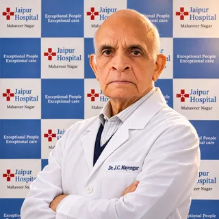
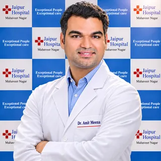
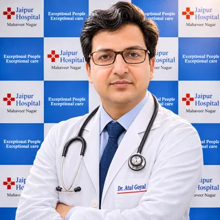
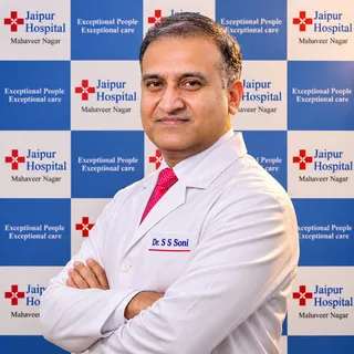
Your orthopaedics doctors
Explore our orthopaedic hospital in Jaipur: every treatment, the doctors and what to expect at your visit.
Frequently asked questions
Questions patients ask about perthes disease treatment
What is Perthes disease treatment?
In short
Perthes disease treatment at Jaipur Hospital aims to keep the femoral head round and inside the socket while the bone heals. Options include observation with activity changes, abduction bracing, or surgery such as femoral or pelvic osteotomy. The choice depends on the child's age, the stage of disease, and how much of the lateral pillar is affected.
In detail
The disease is a self‑limiting avascular necrosis of the femoral head that progresses over two to five years. In children under six with mild involvement (Herring group A), observation and activity modification are usually enough.
For older children or those with lateral pillar B or C disease, containment is needed. Bracing with a Petrie cast or abduction orthosis holds the hip in abduction and internal rotation to maintain coverage.
Surgical containment — femoral varus osteotomy or Salter innominate osteotomy — reorients the bone so the head stays within the acetabulum. Studies show that after age six, operative treatment nearly doubles the chance of a good radiographic outcome compared with non‑operative care.
Both femoral and pelvic osteotomies give similar results in older children. The goal in every case is to prevent femoral head deformity and later osteoarthritis.
A common belief
Many people think surgery is always needed.
What actually happens
In fact, children under six with mild disease often do well with just activity changes and bracing. Surgery is usually reserved for older children or those with more severe lateral pillar involvement.
Your next step at Jaipur Hospital
Your surgeon will review X‑rays and classify the disease using the Herring lateral pillar system. If bracing is chosen, it is worn for 12 to 24 months with regular physiotherapy.
Surgery takes two to three hours under general anaesthesia and needs a three to five night stay. Recovery after osteotomy is three to six months. Typical costs are ₹20,000 to ₹60,000 for bracing and physiotherapy, and ₹1,50,000 to ₹3,00,000 for surgery.
Which specialist should I see for Perthes disease?
In short
You should see a paediatric orthopaedic surgeon — an orthopaedic specialist who focuses on children's bone and joint conditions. At Jaipur Hospital, the orthopaedics department includes surgeons trained in the assessment and containment treatment of Perthes disease.
In detail
Perthes disease is a rare avascular osteonecrosis of the proximal femur usually occurring in children between five and ten years of age.
Its management requires accurate classification using the Herring lateral pillar system at the fragmentation stage, judgement about containment versus observation, and technical skill in femoral varus osteotomy, Salter innominate osteotomy, or triple pelvic osteotomy when surgery is indicated.
The evidence shows that surgical treatment is effective mainly in children over eight years with lateral pillar B or B/C disease, and that both femoral and pelvic osteotomies give similar results in this group.
In younger children with mild disease, non‑operative care with activity modification and bracing is standard. Because the disease is self‑limiting but the final shape of the femoral head determines long‑term hip function, decisions made early affect outcomes decades later. A surgeon who regularly treats this condition can balance the risks of surgery against the risk of deformity.
A common belief
A general orthopaedic surgeon may not see enough Perthes cases to be familiar with the Herring classification nuances or the specific indications for femoral versus pelvic osteotomy.
What actually happens
Paediatric orthopaedic training includes the growth‑plate considerations that are critical here.
Your next step at Jaipur Hospital
Ask your paediatrician or general practitioner for a referral to the orthopaedic OPD at Jaipur Hospital. Bring any previous X‑rays. The surgeon will examine your child, order fresh radiographs if needed, and discuss whether observation, bracing, or surgery is appropriate. Typical non‑surgical care costs ₹20,000 to ₹60,000; surgery ranges from ₹1,50,000 to ₹3,00,000.
What treatment options are available for Perthes disease?
In short
Treatment options at Jaipur Hospital are observation with activity modification, abduction bracing (Petrie casts or orthosis), and surgical containment with femoral or pelvic osteotomy. The choice depends on the child's age at onset, the Herring lateral pillar group, and the degree of femoral head involvement.
In detail
For children under six with Herring group A disease, observation and activity restriction are usually sufficient. Containment bracing holds the femoral head in the acetabulum and is worn for 12 to 24 months with physiotherapy.
For children over eight years or those with lateral pillar B or C disease, surgical containment is considered. Femoral varus osteotomy and Salter innominate osteotomy are the standard procedures; both improve head sphericity compared with non‑operative treatment in older children.
Studies show that after age six, operative treatment nearly doubles the chance of a successful radiographic outcome. In severe cases, combined femoral and pelvic osteotomy or triple pelvic osteotomy may be used.
In later stages with established deformity, non‑containment procedures such as valgus extension osteotomy or cheilectomy can relieve symptoms.
The goal in every approach is to prevent femoral head deformity and reduce the risk of early osteoarthritis, which rarely develops before age 50 but depends on final joint congruence.
A common belief
A common belief is that surgery gives a better result for everyone.
What actually happens
Evidence shows that under age six, surgery does not improve femoral head sphericity compared with non‑operative care. Surgery is most beneficial after age six with lateral pillar B or C involvement. The surgeon matches the treatment to the child's specific classification.
Your next step at Jaipur Hospital
Your surgeon will explain the recommended option after classifying the disease. Non‑surgical care is outpatient and costs ₹20,000 to ₹60,000 for bracing and physiotherapy.
Surgery takes two to three hours under general anaesthesia, needs a three to five night stay, and recovery is three to six months. Surgical costs typically range from ₹1,50,000 to ₹3,00,000. Regular follow‑up X‑rays track healing over the two to five year disease course.
What can I safely do at home to manage symptoms of Perthes disease?
In short
At home, you can help by encouraging rest during painful episodes, using crutches if the surgeon advises, and doing the prescribed physiotherapy exercises daily. At Jaipur Hospital, the team teaches parents how to assist with gentle range-of-motion movements, especially abduction and internal rotation, to keep the hip mobile without overloading it.
In detail
Evidence suggests rest for five to seven days with anti-inflammatory medication may minimise symptoms during a flare. If motion does not return, traction or further assessment may be needed. Once maximum motion is regained, containment therapy is considered for groups with poor prognosis.
Physiotherapy aims to maintain muscle strength and prevent contractures. Avoid high-impact activities such as running, jumping, and contact sports while the femoral head is healing. Swimming and cycling are often allowed because they load the joint less.
The surgeon will give specific guidance based on the stage and classification. A firm mattress and proper positioning during sleep can improve comfort. Regular follow-up X-rays track the femoral head shape.
A common belief
Some families stop all movement to protect the hip.
What actually happens
That can cause stiffness and muscle loss. The right balance is protected weight-bearing with regular motion exercises guided by a physiotherapist.
Your next step at Jaipur Hospital
Ask the physiotherapist at Jaipur Hospital for a written home exercise plan. Review it at each follow-up visit. Keep a symptom diary to share with the surgeon. Call 0141-2551500 or 7414 001 001 to arrange a physiotherapy session.
Is this change part of normal growth or does it need treatment for Perthes disease?
In short
A limp, hip or knee pain, and limited hip movement — especially reduced abduction and internal rotation — are not normal growing pains. These signs suggest Perthes disease, an avascular necrosis of the femoral head that usually appears between ages three and twelve. Jaipur Hospital evaluates each child with clinical examination and radiographs to decide whether observation, bracing, or surgery is required.
In detail
Perthes disease is a rare avascular osteonecrosis of the proximal femur, not a variant of normal growth. The cause of the ischaemia remains unknown, and no preventive strategy exists.
Clinical signs include hip and knee pain, reluctance to move, and restricted range of motion, particularly in abduction and internal rotation. Radiographs are classified using the Herring lateral pillar system, which has high prognostic value and reproducibility.
Age at onset and lateral pillar group are the two main predictors of long‑term outcome. Children under six with mild lateral pillar involvement (Group A) may be managed with observation and activity modification.
Older children or those with Group B or C disease often need containment bracing or osteotomy to preserve femoral head sphericity and hip congruence.
A common belief
Perthes disease is avascular necrosis of the femoral head, not a growth variant.
What actually happens
It follows a defined course over years. The Herring classification on X‑ray tells us the severity. Children under six with mild changes may only need observation, but older children or those with more lateral pillar loss often require bracing or surgery to keep the femoral head round and well seated.
Your next step at Jaipur Hospital
If your child has a persistent limp or hip discomfort lasting more than a week, schedule an OPD visit. The surgeon will take a focused history, examine hip range of motion, and order standing pelvis and frog‑leg lateral X‑rays. Classification and treatment planning usually happen in the same visit.
How will Perthes disease treatment affect my walking, movement, or independence?
In short
During active treatment, walking and independence change temporarily. Non‑surgical care with bracing or activity modification limits high‑impact play for eighteen to twenty‑four months but allows protected weight‑bearing. After femoral or pelvic osteotomy, your child uses crutches or a walker for six to twelve weeks, then progresses to full weight‑bearing over three to six months. Jaipur Hospital guides each phase with physiotherapy to restore motion and strength.
In detail
The disease itself causes hip pain, stiffness, and limited abduction and internal rotation. Non‑surgical containment aims to preserve hip motion while the femoral head remodels over eighteen to twenty‑four months. Bracing restricts abduction but permits walking with aids.
Surgical containment (femoral varus osteotomy or Salter innominate osteotomy) takes two to three hours under general anaesthesia with a three to five night stay. Post‑operative immobilisation and protected weight‑bearing last six to twelve weeks, followed by progressive strengthening for three to six months.
Studies show surgical treatment improves femoral head sphericity in children over six. Long‑term outcome in adulthood is usually good, with osteoarthritis rarely before fifty years. The risk of later arthritis depends on final joint congruence. Physiotherapy throughout treatment maintains range of motion and gait pattern.
A common belief
Parents often fear long-term disability.
What actually happens
The evidence shows most children regain normal function. The disease itself stiffens the hip; treatment — whether brace or osteotomy — is designed to protect the femoral head so it heals round. Physiotherapy keeps muscles active during the restricted phase. Long‑term studies report good adult outcomes, with arthritis uncommon before fifty.
Your next step at Jaipur Hospital
Before surgery, the physiotherapy team teaches crutch walking and home exercises. After discharge, weekly outpatient physiotherapy continues for six to eight weeks, then tapers as strength returns. School and sport restrictions are explained at each stage. Most children resume normal daily activities by three months and unrestricted play by six months after osteotomy.
Which symptoms or changes should prompt me to seek medical advice for Perthes disease?
In short
You should seek medical advice if your child develops a limp, complains of hip or knee pain, or shows reluctance to walk or run. Stiffness in the hip, especially reduced abduction and internal rotation, is a key sign. These symptoms often appear gradually and may come and go, but they should not be ignored.
In detail
Clinical signs of Perthes disease include hip and knee pain, reluctance to move, and limited range of motion, particularly in abduction and internal rotation of the hip joint.
The pain may be felt in the thigh or knee rather than the hip itself, which can delay recognition. A limp that worsens with activity and improves with rest is typical. Over time, muscle wasting around the hip and a fixed flexion contracture may develop.
The disease usually appears between ages three and twelve, with the highest rate at five to seven years. Boys are affected three to five times more often than girls. Bilateral involvement occurs in 10 to 24 percent of cases.
Early evaluation allows classification by the Herring system during the fragmentation stage, which guides treatment decisions and improves long‑term outcomes.
A common belief
Parents often mistake the pain for a knee problem or growing pains because the child points to the knee.
What actually happens
Also, the limp may be intermittent. Do not wait for the limp to become constant; early X‑ray and classification change the treatment options.
Your next step at Jaipur Hospital
If you notice any of these signs, bring your child to the orthopaedic OPD at Jaipur Hospital. The doctor will take a history, examine the hip, and order an X‑ray. Further imaging such as MRI may be advised at an outside centre. Early assessment means treatment can start during the optimal window.
What will the doctor ask and check at the first visit for Perthes disease?
In short
At the first visit, the doctor will ask when the limp or pain started, whether it worsens with activity, and if there is any history of injury or fever. They will examine your child's gait, measure hip range of motion — especially abduction and internal rotation — check for muscle wasting, and assess leg length. An X‑ray of the pelvis and hip will be ordered.
In detail
The clinical signs of Perthes disease are hip and knee pain, reluctance to move, and limited range of motion, particularly in abduction and internal rotation. The doctor will look for a Trendelenburg gait, fixed flexion contracture, and shortening of the affected limb.
They will ask about the child's age at onset, because age over six years and lateral pillar B or C classification are the main predictors of outcome.
The Herring classification, based on the height of the lateral pillar at the fragmentation stage, has high prognostic value and good reproducibility. The Salter‑Thompson and Catterall systems may also be mentioned, but Herring is the most widely used today.
The doctor will also note risk factors such as overweight, female sex, and bilateral involvement, which are linked to poorer prognosis. If the hip is irritable, a short period of rest and NSAIDs may be advised before formal containment therapy begins.
A common belief
Some parents expect an MRI straight away.
What actually happens
X‑ray is the first and most important test because the Herring classification — which guides treatment — is based on X‑ray appearance at the fragmentation stage. MRI is added only if the X‑ray is unclear or hinge abduction is suspected.
Your next step at Jaipur Hospital
Bring any previous imaging and a list of medicines. The X‑ray is done in‑house at Jaipur Hospital. If MRI is needed for early staging or to assess hinge abduction, the surgeon will refer you to an outside imaging centre and review the results.
After classification, the surgeon will explain whether observation, bracing (12 to 24 months), or surgery (2 to 3 hours, 3 to 5 night stay) is recommended.
Can Perthes disease improve without a procedure or operation?
In short
Yes, Perthes disease can improve without surgery in many children. At Jaipur Hospital, the orthopaedic team assesses age, X-ray findings and hip movement to decide if non-surgical care is suitable. Children under six years with mild changes often do well with observation and activity modification alone.
In detail
Evidence shows that in children younger than six years, operative and non-operative treatments are equally likely to result in a successful radiographic outcome.
For mild cases classified as Herring lateral pillar A, or Catterall grades I and II, the femoral head often remodels well with time. The disease is self-limiting and progresses through defined stages over two to five years.
Non-surgical management involves protecting the hip from excessive load while maintaining movement. This may include rest during painful phases, temporary use of crutches, and physiotherapy to preserve abduction and rotation. The aim is to keep the femoral head within the acetabulum during the healing phase. Regular clinical and X-ray reviews track the shape of the femoral head and the congruence of the joint.
A common belief
A common belief is that all cases need bracing or surgery.
What actually happens
Evidence shows that for younger children with mild disease, observation gives results similar to surgery. The femoral head often remodels naturally over two to five years.
Your next step at Jaipur Hospital
If your child has a limp or hip discomfort, book an OPD consultation at Jaipur Hospital. The surgeon will examine the hip, review X-rays and explain whether observation is appropriate. Follow-up visits are typically every three to six months during the active phase.
Are there foods, activities, or habits I should change for Perthes disease?
In short
There is no specific diet that alters the course of Perthes disease, but maintaining a healthy weight is important. At Jaipur Hospital, the orthopaedic team advises that overweight is a clinical factor linked to poorer prognosis. Activities involving running, jumping and contact sports should be avoided during the active phase. Swimming and cycling may be allowed once the surgeon confirms adequate containment.
In detail
The cause of the femoral head ischaemia remains unknown, so no nutritional supplement or food has been shown to prevent or reverse the condition. A balanced diet with adequate calcium and vitamin D supports general bone health.
The main habit change is consistent adherence to weight-bearing restrictions and brace wear if prescribed. Smoking in the household should be avoided as second-hand smoke may impair bone healing.
Regular school attendance is encouraged with modifications such as extra time between classes, elevator access and exemption from physical education. The disease typically lasts two to five years, so sustainable routines are better than short-term strictness that cannot be maintained.
A common belief
Parents often look for a supplement or superfood to fix the problem.
What actually happens
The evidence shows the cause is unknown and no food alters the course. The real influence is mechanical: protecting the femoral head from excessive load while it remodels.
Your next step at Jaipur Hospital
Discuss your child's current diet, sports and school routine with the surgeon and physiotherapist. They will give specific activity clearance based on the latest X-rays.
FAQ library
Questions and answers about perthes disease treatment
When you contact us
Get a clear plan for your child
Contact Jaipur Hospital for an orthopaedic OPD consultation, scan review, plain explanation of options, a cost estimate, cashless insurance help and follow-up care.
- A consultation with an orthopaedic surgeon in the OPD
- Your scans and reports reviewed, with tests done in-house if you need them (laboratory, X-ray and imaging)
- Your options explained in plain words, including whether you need surgery at all
- A cost estimate before you decide
- Help with cashless treatment under MAA Yojana, various government health care schemes and empanelled insurers and TPAs
- Follow-up visits after your treatment
Overview
Perthes Disease Treatment: what to know
What is Perthes disease
- 10 to 24 percent Both hips involved
- 0.2 to 19.1 per 100,000 Annual incidence
Perthes disease is a rare condition in which the blood supply to the ball of the thigh bone (femoral head) is temporarily interrupted. Without blood, the bone tissue dies, a process called avascular necrosis. The body then removes the dead bone and replaces it with new bone over a period of two to five years.
This disease usually affects children between five and ten years of age. The highest rate of occurrence is between five and seven years. Boys are affected three to five times as often as girls. In about 10 to 24 percent of children both hips are involved.
The annual incidence among children under 15 years varies widely, from 0.2 to 19.1 per 100,000. The exact cause of the loss of blood supply is not known.
Symptoms and warning signs that need urgent care the same day
- Pain not settling with rest
- Cannot bear weight
- Hot hip with fever
The most common signs are:
- Hip pain
- Knee pain
- Reluctance to move the leg
- Limited range of motion, especially in abduction and internal rotation of the hip
A child may limp or complain of pain in the thigh or knee rather than the hip itself. Pain that does not settle with rest, sudden inability to bear weight on the leg, or a hip that feels hot and is accompanied by fever should be assessed on the same day. These features may indicate an infection or another urgent problem that needs immediate attention.
Causes and who is at higher risk
- Age over six years
- Clinical factor of poor prognosis
- Overweight
- Associated with worse outlook
- Female sex
- Associated with worse outlook
- Limited hip abduction
- Associated with worse outlook
- Family history
- Present in 8 to 12 percent
- Both hips involved
- 10 to 24 percent, poorer prognosis
The cause of the ischaemia that leads to necrosis of the femoral head remains unknown. Current knowledge does not allow effective preventive strategies. Several factors are linked to a higher risk or a poorer prognosis. Age at onset exceeding six years is a clinical factor of poor prognosis.
Overweight, female sex, and lack of hip abduction are also associated with a worse outlook. Race and latitude influence incidence: the disease is more common at higher latitudes and in certain racial groups. A family history is present in approximately 8 to 12 percent of patients. Bilateral involvement occurs in 10 to 24 percent of cases and carries a poorer prognosis.
How Perthes disease is diagnosed: examination and tests
The doctor will examine the hip for pain, limp, and restriction of movement, especially abduction and internal rotation. Plain X-rays are the main imaging tool. The Herring classification, based on the height of the lateral pillar of the femoral head during the fragmentation stage, is the most widely used system and has good prognostic value.
Herring Group A means the lateral pillar is intact. Group B means height loss of less than 50 percent. Group C means height loss greater than 50 percent. The Catterall classification grades the extent of the epiphyseal lesion from Type I to IV. The Salter-Thompson classification uses the subchondral fracture seen on early X-rays.
MRI is increasingly used and can show early changes that correlate with radiographic classifications. Advanced imaging has also improved the assessment of hinge abduction, although its exact definition remains controversial. The role of imaging in management is rapidly evolving. New techniques may eventually allow earlier prognosis and treatment.
Treatment options without surgery and when surgery is advised for Perthes disease
The goal of treatment is to prevent femoral head deformity. In children under six years, observation and activity modification are often recommended because surgical treatment did not alter femoral head sphericity in this age group. For mild cases (Herring Group A) or children under six, non-surgical management with activity modification and sometimes bracing is used.
Non-surgical treatment typically lasts 12 to 24 months with activity modification for 18 to 24 months. Containment bracing such as Petrie casts or an abduction orthosis keeps the femoral head within the acetabulum. Surgery is now performed considerably less often than in the past.
It is effective mainly in patients with lateral pillar Group B or B/C disease with onset after eight years of age. In children six years and older, operative treatment is nearly twice as likely to result in a successful radiographic outcome compared with non-operative treatment.
Surgical options include femoral varus osteotomy, Salter innominate osteotomy, or triple pelvic osteotomy to improve containment. In severe forms, combining femoral and pelvic osteotomies may be preferable. For skeletally mature patients with established deformity, procedures such as cheilectomy or hip replacement may be considered.
The choice between femoral and pelvic osteotomy varies by region; femoral osteotomy is more common in Europe, Asia and Africa. Your surgeon will advise which approach suits your child based on age, classification, and hip motion.
Preparing for Perthes disease surgery
Do
- Bring X-rays and MRI reports
- Bring regular medicine prescriptions
- Follow fasting instructions
- Pack loose clothing and favourite toy
When your surgeon recommends an osteotomy for Perthes disease, the first step is a detailed assessment in the OPD. The doctor will review your child's X-rays and any MRI reports from an imaging centre to confirm the Herring lateral pillar group and measure the range of motion, especially abduction and internal rotation.
Blood tests such as a complete blood count, bleeding and clotting times, and a basic metabolic panel are usually done a few days before admission. If your child takes any regular medicines, bring the prescriptions so the anaesthesia team can advise which to continue or pause.
Fasting instructions are given the evening before surgery: typically no solid food for six hours and clear fluids up to two hours before the scheduled time. Pack comfortable loose clothing, a favourite toy or book, and any orthopaedic aids your child already uses. An attendant should plan to stay for the 3 to 5 nights of the hospital stay.
How Perthes disease surgery is done
- General anaesthesia Operation takes 2 to 3 hours
- Choose containment procedure Based on age, Herring group, and deformity
- Femoral varus osteotomy Cut upper femur, angle neck, fix with plate and screws
- Salter innominate osteotomy Reorient pelvic bone for better coverage
- Combined or triple pelvic osteotomy For more severe cases
- Intra-operative imaging confirms correction Before wound is closed
The operation is performed under general anaesthesia and takes about 2 to 3 hours. The surgical team uses one of two main containment procedures, chosen according to your child's age, the Herring lateral pillar classification, and the degree of femoral head deformity.
A femoral varus osteotomy (FVO) involves a controlled cut in the upper femur, angling the femoral neck so the head sits deeper in the acetabulum; the bone is held with a plate and screws. A Salter innominate osteotomy (SIO) reorients the pelvic bone to improve coverage of the femoral head.
In more severe cases, the surgeon may combine both procedures or perform a triple pelvic osteotomy (TPO) to achieve stable containment. Intra-operative imaging confirms the correction before the wound is closed. After surgery, a hip spica cast or an abduction brace is often applied to protect the osteotomy while the bone heals.
Risks and possible complications of Perthes disease surgery
- Superficial wound infection
- Common early complication
- Temporary nerve irritation
- Numbness or weakness
- Delayed bone healing (non-union)
- May need further surgery
- Hip stiffness
- From long cast or brace wear
- Leg length discrepancy
- Late effect, especially after femoral osteotomy
Every operation carries some risk. In studies of children undergoing femoral or pelvic osteotomy for Perthes disease, the most common early complications include superficial wound infection, temporary nerve irritation causing numbness or weakness, and delayed bone healing (non-union) which may need further surgery.
Stiffness of the hip joint can occur if the cast or brace is worn for a long period; physiotherapy helps minimise this. Leg length discrepancy is a known late effect, particularly after femoral osteotomy, and may require a shoe raise or later equalisation surgery.
In long-term follow-up of patients who eventually needed total hip replacement for Perthes sequelae, intra-operative fracture occurred in 11% when standard femoral stems were used, aseptic loosening in 5%, sciatic nerve palsy in 3%, and heterotopic ossification in 2%.
All patients who developed sciatic nerve palsy after hip replacement had a history of previous hip surgery, and the average limb lengthening in those cases was 1.9 cm. Your surgeon will discuss which of these risks apply most to your child's situation.
Recovery and aftercare after Perthes disease surgery
- Hospital stay 3 to 5 nights
- Weight-bearing restricted First 6 weeks
- Cast or brace removed About 6 to 8 weeks
- Gradual return to full activity 3 to 6 months
- Follow-up visits 2 weeks, 6 weeks, 3 months, 6 months, then yearly
After the operation, your child stays in the hospital for 3 to 5 nights. Pain is managed with regular medication, and the nursing team monitors the cast or brace for pressure areas. Before discharge, the physiotherapist teaches you how to help your child move safely, use walking aids if needed, and perform gentle exercises at home.
For the first 6 weeks, weight-bearing is usually restricted; the exact timeline depends on the osteotomy type and bone quality. The cast or brace remains in place for about 6 to 8 weeks, after which X-rays confirm healing. Gradual return to full activity takes 3 to 6 months.
Follow-up visits are scheduled at 2 weeks, 6 weeks, 3 months, and 6 months, then yearly until skeletal maturity. Contact the hospital if you notice increasing pain, swelling, redness or discharge at the wound, fever, numbness or colour change in the toes, or if the cast becomes loose or cracked.
Results of Perthes disease treatment
The goal of treatment is to prevent femoral head deformity and maintain a congruent hip joint. In children younger than 6 years, operative and non-operative treatments are equally likely to result in a successful radiographic outcome. For children aged 6 years and older, operative treatment is nearly twice as likely to achieve a good radiographic outcome compared with non-operative care.
Femoral varus osteotomy and Salter innominate osteotomy provide good outcomes in moderate forms of the disease. In severe forms, combining the two techniques or performing a triple pelvic osteotomy seems preferable. Age at onset and the Herring lateral pillar classification are the two main predictors of long-term outcome.
Patients between 5 and 7 years old have a better prognosis than older children, while adolescence at onset carries a poor prognosis. Bilateral involvement also predicts a poorer outcome. The risk of hip osteoarthritis in adulthood depends chiefly on the final degree of joint incongruence; osteoarthritis rarely develops before 50 years of age.
In studies of total hip replacement for Perthes sequelae, all reported a significant improvement in hip function. The mean age at hip replacement was 45.7 years with a mean follow-up of 8.4 years, and 16 revisions occurred at a mean of 7.5 years.
Cost and insurance for Perthes disease treatment in Jaipur
The typical cost range for non-surgical management with bracing and physiotherapy is ₹20,000 to ₹60,000. Surgical treatment with femoral or pelvic osteotomy typically costs ₹1,50,000 to ₹3,00,000. The final amount depends on the type of osteotomy, the implants used, the length of hospital stay, and whether both hips are treated.
Jaipur Hospital is empanelled under MAA Yojana, various government health care schemes, and a panel of private insurers and TPAs. Bring your scheme card and insurance documents at admission so the accounts team can verify coverage and process the claim.
The hospital has an in-house pharmacy and diagnostic laboratory; X-rays are done on site, while MRI or CT scans are arranged at an external imaging centre and billed separately.