

Plastic Surgery · Jaipur Hospital
Peripheral Nerve Surgery in Jaipur
Numbness, weakness or pain after nerve injury — we assess, explain options and plan your treatment.
- Serving Jaipur since 1991 (35 years)
- NABH accredited
- 24×7 emergency & ICU
- Cashless: RGHS, MAA Yojana, CGHS + 22 insurers
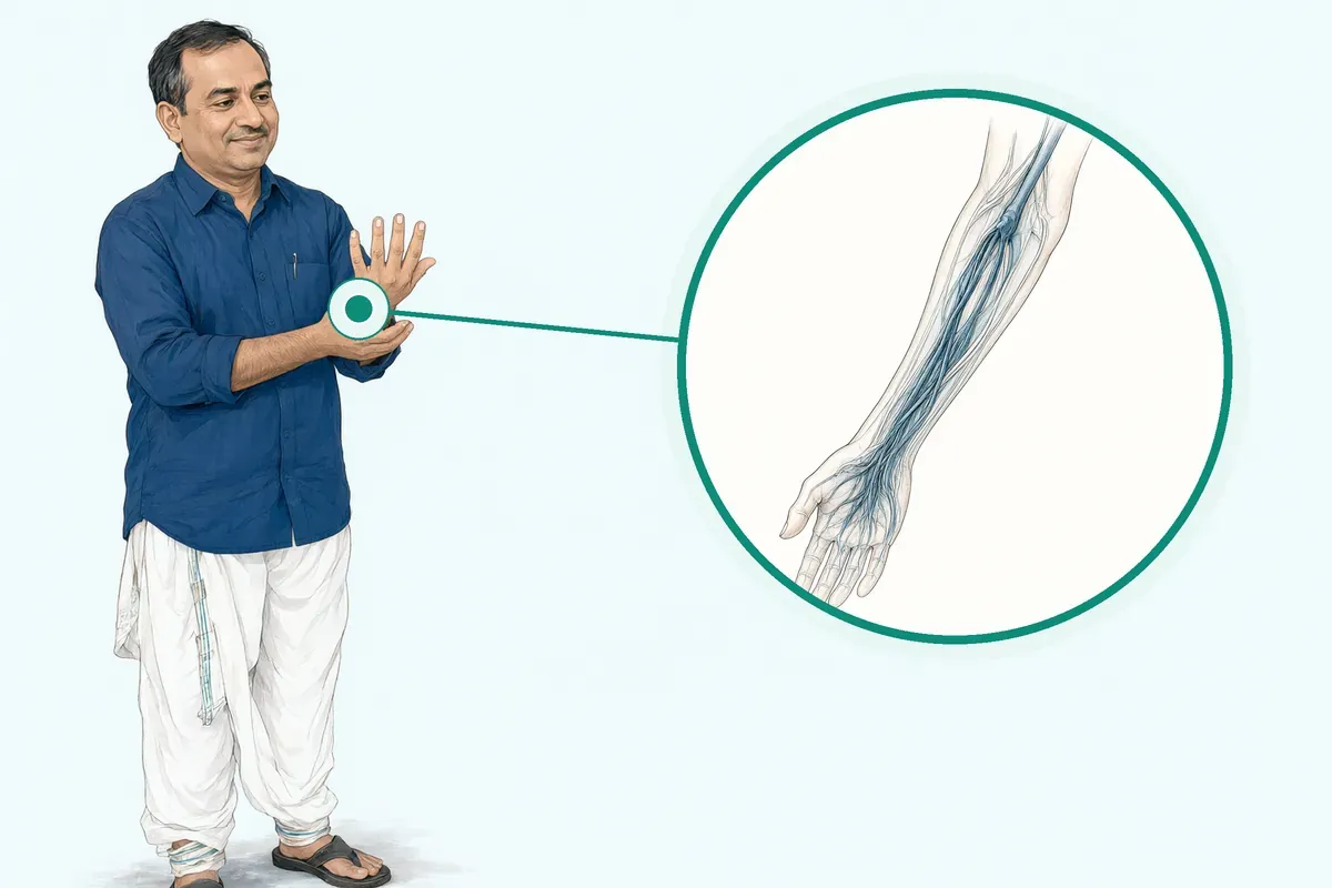
- Operation time
- 1 to 4 hours
- Anaesthesia
- General or regional anaesthesia
- Recovery
- Nerve regeneration 1 mm/daytherapy 6-18 months
- Price
- ₹50,000 to ₹3,50,000typical range in India; ask us for an estimate
Nerve injury disrupts daily life
Loss of feeling, strength or control after an injury
A cut, crush or stretch can damage nerves in your arm or leg. You may feel numbness, tingling, weakness or sharp pain. Simple tasks become hard. Work and sleep suffer. Without timely care, muscles waste and recovery gets harder.
-
Numbness or tingling
Parts of your hand, foot or limb feel dead or prickly.
-
Weak grip or foot drop
You cannot hold things or your foot drags when walking.
-
Sharp or burning pain
Pain shoots along the nerve path, often worse at night.
-
Muscle wasting
The limb looks thinner as muscles shrink from disuse.
-
Daily tasks affected
Buttoning a shirt, typing or climbing stairs becomes difficult.
Peripheral nerves lack the bony protection of the brain and spinal cord, making them easy to damage.
Why Jaipur Hospital
You worry that the weakness will not improve, that pain will keep you awake, and that time off work will stretch your finances.
Your care plan
Your first steps with us
- 1 Call or visit OPD for a surgeon consultation.
- 2 Get scans and nerve tests done in-house.
- 3 Review options and receive a cost estimate.
- 4 Have surgery and start guided physiotherapy.
How the operation is done
Surgery uses a microscope to free, join or bridge the damaged nerve. Anaesthesia is general or regional. The procedure takes one to four hours. Stay ranges from daycare to three nights.
-
Neurolysis / external neurolysis
Releases the nerve from scar tissue or tight fascia compressing it.
-
Direct nerve repair
Microsurgical stitching of cut nerve ends, usually within 72 hours for clean cuts.
-
Nerve graft (sural / MABC)
Uses a sensory nerve from your leg or processed donor nerve to bridge gaps over 2 cm.
-
Nerve transfer
Connects a working donor nerve to a more important paralysed target for faster reinnervation.
Why patients trust us
- 1991 Serving Jaipur for 35 years
- NABH Accredited hospital
- 1 Plastic Surgery consultants
- 24×7 Emergency, ICU and ambulance
- 25 Cashless schemes and insurers
What research shows about nerve surgery outcomes
-
1 to 2 mm per day
Axons regenerate at this rate after repair; no therapy speeds it up.
-
12 to 18 months
Accepted window for muscle reinnervation before irreversible end-plate degeneration.
-
Less than half
Fewer than 50% of patients regain good to excellent motor or sensory function after repair.
If it is left untreated
Without surgery, cut or compressed nerves rarely heal on their own. Muscles waste away, joints stiffen, and chronic neuropathic pain can set in. The longer the delay, the lower the chance of useful recovery.
Life after treatment
Most people see gradual return of sensation and strength over many months with dedicated therapy.
-
Sensation returns
Numb areas usually regain feeling, starting near the injury and moving outward.
-
Strength improves
Weak muscles typically get stronger as nerves reconnect, though full power may not return.
-
Pain reduces
Neuropathic pain often lessens once the nerve is decompressed or repaired.
-
Daily function
Most people manage self-care and light work again with continued exercises.
Frequently asked questions
Questions patients ask about peripheral nerve surgery
Which problems are assessed under peripheral nerve surgery?
In short
Peripheral nerve surgery at Jaipur Hospital assesses injuries and conditions affecting nerves in the upper and lower limbs. These include nerve compression, sharp cuts, stretch injuries, and gaps caused by trauma or previous operations. The surgical team evaluates the type and severity of nerve damage to plan appropriate treatment.
In detail
Peripheral nerve injuries (PNI) can have several etiologies such as trauma and iatrogenic interventions leading to loss of structure or function.
Injuries are classified by severity: neurapraxia involves demyelination without axon loss; axonotmesis adds axonal lesion with preserved connective tissue; neurotmesis is complete transection of axons and connective tissue. The majority of peripheral nerve injuries occur in the upper limb from traumatic causes.
Common problems include compressive neuropathies, lacerations, stretch injuries, and nerve gaps requiring reconstruction. The surgical team uses clinical examination and diagnostic tests to determine the injury grade and plan management.
A common belief
Many people think nerve surgery is only for cut nerves.
What actually happens
In fact, compressed nerves, stretch injuries, and old scars causing tethering are also assessed and treated surgically when conservative care fails.
Your next step at Jaipur Hospital
Your surgeon will review your history, examine nerve function, and may order nerve conduction studies or imaging. Based on findings, they will discuss whether surgery is needed and which technique suits your injury.
How will the doctor decide which tests I need?
In short
The surgeon at Jaipur Hospital decides on tests based on your clinical examination findings, the suspected nerve and level of injury, and the time since onset. If the injury is recent, the surgeon may rely on clinical localisation alone and plan early exploration. If it has been more than three to six weeks, nerve conduction studies and electromyography are usually requested to confirm the level and severity. Imaging such as MRI or ultrasound is added when a compressive lesion, neuroma, or soft tissue tumour is suspected.
In detail
Clinical examination maps the sensory and motor deficit to a specific nerve. Nerve conduction studies assess motor and sensory conduction across segments and can identify conduction block. Electromyography detects fibrillations and positive sharp waves in denervated muscle, which appear three to six weeks after injury.
The surgeon correlates these findings with the clinical picture. For example, a suspected carpal tunnel syndrome may only need nerve studies, while a brachial plexus injury typically needs MRI of the plexus and serial EMGs. The choice is individualised; there is no fixed panel of tests for every patient.
A common belief
Some patients expect a standard set of scans for every nerve problem.
What actually happens
But a wrist compression needs different tests than a brachial plexus injury. The surgeon tailors the investigations to your specific clinical picture to avoid unnecessary tests and delays.
Your next step at Jaipur Hospital
The surgeon will explain which tests are needed and why during the consultation. Some tests can be done in-house at the hospital's diagnostic laboratory. Others may be scheduled for a later date. Results are usually available within a few days. The surgeon will then review them with you and finalise the treatment plan.
How soon should I expect improvement after peripheral nerve surgery?
In short
Jaipur Hospital advises that nerve regeneration proceeds at about one millimetre per day, so visible recovery takes months. Sensory return may be noticed earlier, but useful motor function often needs six to eighteen months of dedicated therapy after surgery.
In detail
The evidence shows axonal growth averages one to two millimetres daily. A wrist-level injury requires roughly one hundred days for axons to reach hand muscles; a brachial plexus injury can take two to three years.
Muscle fibrosis begins immediately after denervation and plateaus at four months with sixty to eighty percent volume loss. Motor endplates become unlikely to accept reinnervation after twelve months. Early repair within days of injury gives the highest probability of meaningful recovery.
A common belief
People expect quick results like after fracture fixation.
What actually happens
Nerve regeneration is fundamentally slower, and waiting too long before surgery lets muscles wither beyond repair.
Your next step at Jaipur Hospital
You will start gentle physiotherapy within days of discharge. The surgeon reviews progress at six weeks, three months, and six months with repeat nerve studies. Functional goals are adjusted at each visit.
How can I decide whether to seek advice about brachial plexus reconstruction?
In short
You should seek advice about brachial plexus reconstruction if you have weakness, numbness or pain in the shoulder, arm or hand after a traction injury, fall or road accident. Jaipur Hospital offers clinical examination and surgical exploration to assess the injury. Early evaluation helps determine if surgery can improve recovery.
In detail
Brachial plexus injuries often involve nerve root avulsion or severe stretching that does not heal on its own. Evidence shows that acute repair leads to better functional restoration and delays from a wait-and-see approach can be costly.
The surgical team uses clinical examination and, when needed, surgical exploration because no non-invasive test can diagnose severity in the first six weeks. Nerve conduction studies and EMG are useful only after six weeks.
Reconstruction may involve nerve grafts, nerve transfers or both, depending on which roots are injured and the gap length. The goal is to reinnervate target muscles before irreversible motor end-plate degeneration occurs, typically within 12 to 18 months.
A common belief
Many people wait months hoping the nerve will recover on its own.
What actually happens
Evidence shows that delays beyond three to six months reduce the chance of good recovery because muscles waste away and motor end-plates degenerate.
Your next step at Jaipur Hospital
If you notice persistent weakness or numbness after a shoulder or neck injury, book a consultation at Jaipur Hospital. The surgeon will examine you, review any imaging and discuss whether reconstruction is appropriate. Early referral improves the chance of meaningful recovery.
How will appearance, function, and realistic expectations be discussed?
In short
At Jaipur Hospital, the surgeon discusses appearance, function and realistic expectations during the preoperative consultation. The conversation covers the type of nerve injury, the planned technique such as decompression, repair, graft or transfer, the expected timeline for nerve regeneration at about one millimetre per day, and the likely degree of sensory or motor recovery based on injury level and timing.
In detail
Evidence shows that less than half of patients regain good to excellent motor or sensory function after nerve repair, and each coaptation site results in approximately fifty percent axon loss.
The surgeon explains that recovery is gradual over six to eighteen months with supervised therapy, and that muscle fibrosis begins early after denervation. For proximal injuries such as brachial plexus, distances up to one metre mean two to three years for hand reinnervation.
The discussion includes the possibility of incomplete recovery, the need for secondary procedures such as tendon transfers, and the role of physiotherapy in maintaining joint mobility while awaiting reinnervation. No specific outcome is promised.
A common belief
Many patients expect full normal function.
What actually happens
Evidence shows less than half regain good to excellent function, each repair site loses about fifty percent of axons, and proximal injuries take years. The surgeon will not promise a specific outcome but will outline the probable range.
Your next step at Jaipur Hospital
You will receive a clear verbal summary and have the opportunity to ask questions before signing consent. The team encourages an attendant to be present. Follow-up appointments are scheduled to monitor regeneration clinically and with nerve studies. To start this discussion, book a consultation at Jaipur Hospital.
Which symptoms should prompt me to see a specialist?
In short
You should see a specialist at Jaipur Hospital if you have numbness, tingling, weakness, or loss of movement in an arm or leg after an injury. Neuropathic pain, burning sensations, or a feeling of heaviness in the limb also warrant evaluation. Symptoms that persist beyond a few weeks need expert assessment.
In detail
Peripheral nerve injuries lead to partial or complete loss of sensory, motor, and autonomic functions and neuropathic pain. These changes can cause physical disability and affect quality of life.
Depending on the injury, an extensive array of symptoms and outcomes are possible, contingent on severity, type of trauma, age, and nerves involved.
Common presentations include numbness in a specific nerve distribution, weakness of specific muscle groups, wasting of muscles, and neuropathic pain such as burning or shooting sensations. Autonomic changes like altered sweating or skin colour may also occur.
A common belief
People often ignore gradual numbness or mild weakness, hoping it will resolve.
What actually happens
However, prolonged denervation leads to irreversible muscle atrophy and poorer surgical outcomes. Early specialist review allows timely intervention.
Your next step at Jaipur Hospital
If you notice persistent numbness, weakness, or neuropathic pain after trauma or surgery, do not wait. Early evaluation improves surgical timing and outcomes. Bring any previous reports to your appointment.
How will the results change my care plan?
In short
Test results at Jaipur Hospital directly shape whether you are managed with observation, splinting, physiotherapy, or scheduled for surgery. Nerve conduction studies showing conduction block without axonal loss suggest neurapraxia, which often recovers with conservative care. Electromyography showing active denervation with fibrillations confirms axonotmesis or neurotmesis, which may need surgical repair. MRI or ultrasound showing a neuroma in continuity, a compressive mass, or a nerve gap guides the choice between neurolysis, direct repair, grafting, or nerve transfer.
In detail
The surgeon integrates clinical findings, electrodiagnostic data, and imaging to assign a Sunderland grade to the injury. Grade I (neurapraxia) usually recovers in weeks to months without surgery. Grade II (axonotmesis) may recover spontaneously but is monitored with serial studies.
Grades III to V (neurotmesis) typically require surgical intervention. The timing of surgery also depends on the results: clean sharp cuts are repaired within 72 hours, while stretch injuries or injuries with unclear extent may be observed for three to six months with serial EMGs.
If no clinical or electrophysiological recovery is seen by then, exploration is indicated. The specific technique — neurolysis, epineurial repair, fascicular repair, nerve graft, or nerve transfer — is decided by the intraoperative findings of nerve continuity, gap length, and fascicle matching.
A common belief
Patients often think a normal MRI means no surgery.
What actually happens
But MRI cannot see microscopic axonal loss. EMG and nerve studies are the functional tests that show whether axons are regenerating. The surgeon combines all pieces of information, not just one scan, to decide.
Your next step at Jaipur Hospital
After your tests are done, the surgeon will discuss the results with you and explain the recommended path. This may be a trial of therapy with repeat testing at six to eight weeks, or a date for surgery. The plan is always individualised.
What should I do if symptoms persist or worsen after peripheral nerve treatment?
In short
If symptoms persist or worsen after peripheral nerve treatment, contact your surgeon at Jaipur Hospital promptly. New or increasing weakness, numbness, pain, swelling, redness, or wound discharge should be reported without delay so that early intervention can be arranged.
In detail
Persistent symptoms may indicate incomplete regeneration, neuroma formation, scar tethering, or graft failure. Worsening weakness or new sensory loss can signal compression at the repair site or progression of the original injury.
Infection presents with increasing pain, swelling, warmth, or discharge and needs antibiotics or drainage. A painful lump at the scar may be a neuroma and can be assessed with ultrasound or nerve studies.
Your surgeon may order repeat nerve conduction studies, imaging, or clinical examination to identify the cause. Depending on findings, options include further therapy, medication adjustment, neurolysis, revision repair, or nerve transfer. Early reporting improves the chance of a successful secondary procedure.
A common belief
Some patients hope symptoms will settle on their own.
What actually happens
Delaying review can allow scar tissue to mature or muscle to fibrose, making later correction harder. Early action preserves options.
Your next step at Jaipur Hospital
Keep the emergency and OPD numbers handy. Do not wait for the next scheduled visit if something feels wrong.
How can I decide whether to seek advice about nerve grafting and nerve transfer?
In short
Nerve grafting and nerve transfer are surgical options when a peripheral nerve is cut or has a gap that cannot be repaired directly. Jaipur Hospital provides assessment for these procedures. You should seek advice if you have a sharp nerve laceration, a nerve gap after trauma, or persistent weakness despite conservative care.
In detail
Direct repair is possible only when nerve ends can be joined without tension, usually within 72 hours for clean cuts. If a gap exceeds two centimetres, an autograft (often sural nerve) or processed allograft is used.
Nerve transfers redirect a working donor nerve to a more critical paralysed target, offering faster reinnervation for proximal injuries. Evidence shows that at each coaptation site about 50% of axons cross; with a graft there are two sites, so roughly 25% may regenerate across.
Transfers avoid one coaptation and shorten the distance. The choice depends on injury level, gap length, donor availability, and time since injury. Early referral, ideally within three to six months, preserves muscle targets.
A common belief
People often think any nerve cut can just be stitched.
What actually happens
But tension at the repair site causes scarring and failure. Grafts add a second repair site, so fewer axons get through. Transfers can be better for high injuries where the distance to the muscle is long.
Your next step at Jaipur Hospital
If you have a diagnosed nerve cut, a gap on imaging, or no recovery after three months of therapy, request a surgical opinion. Bring prior reports. The team will explain which technique suits your injury.
What qualifications and experience should the treating clinician have for this procedure?
In short
At Jaipur Hospital, peripheral nerve surgery is performed by surgeons with formal training in microsurgical nerve reconstruction. The treating clinician should hold a recognised postgraduate qualification in plastic surgery, orthopaedics or neurosurgery, have completed fellowship or dedicated training in peripheral nerve and brachial plexus surgery, and regularly perform nerve decompression, repair, grafting and transfer procedures in a multidisciplinary setting with physiotherapy support.
In detail
Nerve surgery requires operating microscope expertise, fine suture technique, and knowledge of nerve anatomy, fascicular topography and donor nerve selection.
The surgeon must be able to decide between direct repair within seventy-two hours for sharp cuts, autograft using sural or medial antebrachial cutaneous nerve for gaps over two centimetres, processed allograft when autograft is unavailable, and nerve transfers such as Oberlin or AIN-to-ulnar for proximal injuries.
The team at Jaipur Hospital works with anaesthetists skilled in regional blocks, neurophysiologists for intraoperative nerve stimulation, and physiotherapists who guide the six to eighteen month rehabilitation protocol. Continuous audit of outcomes and participation in peer review are expected.
A common belief
General surgeons may not have microscope skills or fascicular mapping experience.
What actually happens
Nerve transfers need precise donor selection and coaptation technique. A dedicated nerve surgeon works with neurophysiologists and therapists for the long rehabilitation phase.
Your next step at Jaipur Hospital
You can ask about the surgeon's training, case volume and specific experience with your injury pattern during the OPD consultation. The hospital maintains credentials for all consultants. To meet the team and discuss your case, book an appointment at Jaipur Hospital.
FAQ library
Questions and answers about peripheral nerve surgery
When you contact us
Get a clear plan for your nerve injury
Contact Jaipur Hospital for an OPD consultation, in-house tests, plain-language options, a cost estimate and cashless scheme help.
- A consultation with a plastic surgeon in the OPD
- Your scans and reports reviewed, with tests done in-house if you need them (laboratory, X-ray and imaging)
- Your options explained in plain words, including whether you need surgery at all
- A cost estimate before you decide
- Help with cashless treatment under RGHS, MAA Yojana, CGHS and empanelled insurers and TPAs
- Follow-up visits after your treatment
Overview
Peripheral Nerve Surgery: what to know
What peripheral nerve injury is and how it happens
- Wallerian degeneration starts Within 24 to 48 hours after cut or crush
- Axon growth rate About 1 mm per day in humans
- Wrist injury to hand muscles Roughly 100 mm distance, at least 100 days
- Muscle wasting plateaus After about four months, 60–80% volume lost
- Motor endplate fibrosis Unlikely reinnervation beyond 12 months
Peripheral nerves carry signals between your brain, spinal cord and the rest of your body.
They control:
- Movement
- Feeling
- Automatic functions such as sweating
Unlike the brain and spinal cord, these nerves lie close to the surface with little physical protection, so they are easily damaged.
An injury can stretch, crush or cut a nerve. The mildest form, called neurapraxia, is a temporary block caused by local loss of the nerve's insulating cover (demyelination) without damage to the nerve fibres themselves. A more severe injury, axonotmesis, damages the nerve fibres (axons) but leaves the outer connective tissue intact. The most severe form, neurotmesis, completely cuts through the nerve fibres and their surrounding tissue.
When a nerve is cut or badly crushed, the part of the fibre separated from the cell body breaks down in a process called Wallerian degeneration. This starts within 24 to 48 hours. The distal segment loses its axons and myelin, and immune cells called macrophages arrive to clear the debris.
Schwann cells, which normally support the nerve fibres, change into a repair mode, line up in columns known as bands of Bungner, and release growth factors to guide regenerating axons.
Regeneration is slow. In humans, axons grow at about 1 mm per day. For a wrist-level injury, axons must travel roughly 100 mm to reach hand muscles, taking at least 100 days. More proximal injuries, such as a brachial plexus injury, can require over a metre of regrowth and may take two to three years.
Muscle wasting begins immediately after denervation and plateaus after about four months when 60–80% of muscle volume is lost. Motor endplates become progressively fibrotic, making functional reinnervation unlikely beyond 12 months. Sensory recovery can continue for longer, but earlier repair still gives better outcomes.
Because of these time limits, a rapid intervention is important. Self-repair is not possible for severe or long-distance injuries.
Symptoms of peripheral nerve injury and warning signs that need urgent care the same day
- Growing painful lump near injury site May be a neuroma
- Signs of infection around a wound Redness, warmth, pus, fever
Symptoms depend on which nerve is injured and how severely.
Common features include:
- Partial or complete loss of sensation (numbness, tingling, burning)
- Loss of muscle power or control
- Autonomic changes such as altered sweating or skin colour
Neuropathic pain — often described as shooting, stabbing or electric-shock-like — is frequent and can be disabling.
Warning signs that should prompt same-day medical attention:
- Sudden loss of movement or feeling in a limb after trauma (fall, road accident, cut, crush injury).
- A growing, painful lump near an injury site — this may be a neuroma, a tangled mass of nerve fibres that can form when regenerating axons fail to find their target.
- Progressive weakness or wasting in a limb over days to weeks without an obvious new injury.
- Loss of bladder or bowel control together with limb symptoms — this may indicate a spinal or cauda equina problem needing emergency care.
- Signs of infection (redness, warmth, pus, fever) around a wound that may involve a nerve.
If you notice any of these, go to the emergency department at Jaipur Hospital or the nearest hospital immediately. Early assessment allows your surgeon to decide whether surgical exploration is needed within the optimal window.
Causes of peripheral nerve injury and who is at higher risk
Most peripheral nerve injuries are traumatic. The majority occur in the upper limb.
Common causes include:
- Road traffic accidents
- Falls
- Sports injuries
- Cuts from glass or knives
- Gunshot wounds
- Stretch injuries during difficult childbirth or joint dislocations
Compression over time — for example, from repetitive movements, prolonged pressure, or tight casts — can also damage nerves.
Iatrogenic injury (damage during a medical or surgical procedure) is another recognised cause. This can happen during orthopaedic, vascular, or general surgery, or from poorly placed injections or intravenous lines.
Factors that increase the risk of a poor outcome:
- Delay in diagnosis and repair — each month of denervation reduces the number of axons that can successfully regenerate.
- Proximal injuries (closer to the spinal cord) — longer regeneration distance means more time for muscle fibrosis and motor endplate degeneration.
- Large nerve gaps — gaps greater than 2–3 cm usually cannot be bridged by direct repair and need a graft.
- Age — older patients tend to have slower and less complete regeneration.
- Smoking and uncontrolled diabetes — both impair microvascular blood flow and nerve healing.
- Severe crush or blast injuries — extensive tissue damage creates a hostile environment for regeneration.
Macrophages and Schwann cells play central roles in the natural repair process. After injury, macrophages are recruited to the site, peak around day 7, and switch from a pro-inflammatory (M1) to a pro-repair (M2) phenotype to support debris clearance, angiogenesis and Schwann cell migration. Schwann cells dedifferentiate, clear myelin, form bands of Bungner, and later remyelinate regenerated axons.
Chronic denervation leads to irreversible changes in both Schwann cells and neurons, which is a major reason for poor functional recovery. Misdirection of regenerating axons into wrong endoneurial tubes also limits recovery.
How peripheral nerve injury is diagnosed: examination and tests
- Fibrillations appear late
- Three to six weeks after injury
- No reliable acute test
- First six weeks after injury
- Serial tests map recovery
- Distinguish neurapraxia from axonotmesis
Diagnosis begins with a detailed clinical examination. Your surgeon will test muscle strength, sensation, reflexes, and look for wasting, Tinel's sign (tingling on tapping over the nerve), and autonomic changes. This clinical assessment is the cornerstone of diagnosis, especially in the first six weeks after injury.
Nerve conduction studies (NCS) and electromyography (EMG) are the main electrodiagnostic tests. NCS uses skin-surface electrodes to measure how fast and how well electrical signals travel along motor and sensory nerves. EMG uses a fine needle inserted into muscle to detect abnormal spontaneous activity (fibrillations, positive sharp waves) and to analyse voluntary motor units.
Important timing considerations:
- Fibrillations from denervated muscle may not appear until three to six weeks after injury, depending on how proximal the lesion is.
- There is no non-invasive test that can reliably diagnose the presence or severity of a nerve injury in the first six weeks.
- Serial NCS and EMG over time can map recovery and help distinguish neurapraxia (conduction block only) from axonotmesis (axonal loss).
If there is no clinical or electrophysiological recovery after three to six months, surgical exploration is usually warranted, depending on the level of injury. A "wait-and-see" approach can be costly because acute repair leads to better functional restoration. There remains a great clinical need for accurate acute diagnostics.
At Jaipur Hospital, our neurosurgery and orthopaedics teams work together with the in-house neurophysiology laboratory to perform these tests and plan timely intervention.
Non-surgical treatment options for peripheral nerve injury and when surgery is advised
Releasing nerve from scar tissue or tight fascia
- Neurolysis
Microsurgical stitching of nerve ends, ideally within 72 hours for sharp cuts
- Direct nerve repair
Autograft using sural nerve or medial antebrachial cutaneous nerve, or processed allograft for gaps over 2 cm
- Nerve graft
Connecting a working donor nerve to a more important denervated target (e.g., Oberlin transfer, AIN-to-ulnar)
- Nerve transfer
Not every nerve injury needs surgery. For mild compression or neurapraxia, conservative management is often tried first. This can include:
- Rest and activity modification to avoid further stretch or pressure on the nerve.
- Splinting or bracing to protect the nerve and maintain joint position.
- Physiotherapy to maintain joint range of motion, prevent contractures, and strengthen unaffected muscles.
- Medications for neuropathic pain (such as certain anticonvulsants or antidepressants) — prescribed by your doctor.
- Nerve blocks for temporary pain relief.
Peripheral nerve surgery is typically recommended when conservative treatments such as physical therapy, medication, or splinting have failed to provide relief from symptoms. Surgery is also advised for:
- Complete nerve transection (neurotmesis) — the nerve ends will not reconnect on their own.
- Large nerve gaps where direct repair is impossible without tension.
- Progressive neurological deficit despite conservative care.
- Painful neuroma not responding to other measures.
- Iatrogenic injuries recognised at the time of surgery or soon after.
The choice of surgical technique depends on the specific condition and extent of nerve damage. Options include:
- Neurolysis (external neurolysis): releasing the nerve from scar tissue or tight fascia.
- Direct nerve repair (epineurial or fascicular): microsurgical stitching of the nerve ends, ideally within 72 hours for sharp cuts.
- Nerve graft (autograft using sural nerve or medial antebrachial cutaneous nerve, or processed allograft): used when a gap of more than 2 cm prevents tension-free direct repair.
- Nerve transfer (e.g., Oberlin transfer, AIN-to-ulnar): connecting a working donor nerve to a more important denervated target to achieve faster reinnervation.
Your surgeon will discuss which option suits your injury pattern, the time since injury, and your functional goals. The operation typically takes 1 to 4 hours under general or regional anaesthesia. Hospital stay ranges from daycare to three nights. Nerve regeneration proceeds at about 1 mm per day, and supervised therapy continues for 6 to 18 months.
The typical cost range in India is ₹50,000 to ₹3,50,000; ask us for an estimate based on your specific procedure.
Preparing for peripheral nerve surgery
Do
- Attend pre-anaesthesia check-up
- Bring regular medicines in original packets
Before your operation you will attend a pre-anaesthesia check-up where the team reviews your medical history, current medicines and any allergies. Blood tests, a chest X-ray and an ECG are usually done to confirm you are fit for general or regional anaesthesia.
If you take blood-thinning medicines, diabetes tablets or insulin, your surgeon and anaesthetist will tell you which ones to stop and when to restart them. You must not eat or drink for at least six hours before the scheduled time. Wear loose clothing that can fit over a bulky dressing or splint.
Bring your regular medicines in their original packets, your insurance or scheme cards (RGHS, MAA Yojana, CGHS or private TPA details), and an attendant who can stay with you overnight if admission is needed. The hospital provides 24-hour laboratory, X-ray and ECG services on site, so any last-minute tests can be completed quickly.
How peripheral nerve surgery is done
- Anaesthesia and incision General or regional anaesthesia; incision over injured nerve
- Explore and assess damage Surgeon examines the nerve to decide the technique
- Neurolysis for compression Release nerve from scar tissue or tight fascia
- Nerve transfer for proximal injuries Oberlin transfer or AIN-to-ulnar transfer for faster reinnervation
- Close incision and apply protective splint Sutures or staples; splint protects the repair
The operation takes one to four hours under general or regional anaesthesia. After you are anaesthetised, the surgeon makes an incision over the injured nerve and carefully explores it to assess the damage. The technique used depends on the type and length of the gap.
For a nerve compressed by scar tissue or tight fascia, neurolysis (external neurolysis) releases the nerve. If the nerve is sharply cut and the ends can be brought together without tension within about 72 hours, direct nerve repair with microsurgical stitches (epineurial or fascicular coaptation) is performed.
When the gap is larger than two centimetres, a nerve graft is needed; the sural nerve from the leg or the medial antebrachial cutaneous nerve from the arm are common autograft choices, and a processed allograft is an alternative when donor nerves are limited.
For proximal injuries where the target muscle is far away, a nerve transfer such as an Oberlin transfer or an anterior interosseous nerve to ulnar nerve transfer may be chosen to reinnervate the muscle faster. Your surgeon will explain which option suits your injury.
After the repair, the incision is closed with sutures or staples and a protective splint is applied. The hospital stay ranges from daycare discharge to three nights depending on the complexity.
Risks and possible complications of peripheral nerve surgery
- 50% 50%
- 25% 25%
All operations carry some risk. Infection, bleeding and wound healing problems can occur with any incision. Specific to nerve surgery, the repair site always develops some scarring and fibrosis from sutures and tissue handling, and there is unavoidable size and fascicle mismatch between the nerve ends; these factors can limit regeneration.
In studies, about 50% of the original axons successfully regenerate across a single repair site, and only about 25% cross a nerve graft with two coaptation sites. If regenerating axons do not find their correct endoneurial tubes, they can form a painful neuroma at the repair site or at the donor nerve harvest area.
Donor site morbidity includes long-term sensory loss and a scar where the sural or medial antebrachial cutaneous nerve was taken. Large diameter donor nerves may fibrose internally before axons cross the graft. Processed allografts avoid donor site morbidity but carry significant cost and complexity.
Hollow nerve conduits are generally limited to sensory nerve gaps under three centimetres and have not shown equivalent results to autografts for longer gaps, motor nerves or proximal injuries. Your surgical team will discuss the risks that apply to your specific injury pattern.
Recovery and aftercare after peripheral nerve surgery
After surgery your limb is immobilised in a splint or cast to protect the repair. You will receive pain relief and instructions on wound care before discharge. The first follow-up is usually within one to two weeks for suture removal and wound check.
Nerve regeneration proceeds at about one millimetre per day, so meaningful recovery takes six to eighteen months of dedicated physiotherapy. During this time you will work with the physiotherapy department on joint mobility, sensory re-education and muscle re-training as the nerve grows.
Muscle wasting begins immediately after denervation and plateaus around four months with 60–80% volume loss; motor endplates become unlikely to accept reinnervation after about twelve months due to fibrosis. Sensory recovery can continue for years but earlier repair gives better outcomes.
Contact the hospital promptly if you notice increasing pain, redness, swelling, discharge from the wound, fever, or sudden loss of movement or sensation that was present after surgery. The 24×7 emergency department is available for urgent concerns.
Results of peripheral nerve surgery
In studies, less than half of patients who undergo nerve repair after injury regain good to excellent motor or sensory function. For a primary repair, approximately 50% of the original axons successfully regenerate through the repair site. For a nerve graft with two coaptation sites, about 25% of axons cross the graft.
Additional axonal loss occurs over the distance to the target muscle or sensory organ due to chronic denervation and muscle fibrosis. Proximal injuries such as brachial plexus lesions may require more than two to three years for axons to reach the hand.
Early repair leads to better functional restoration; delays of more than four months can reduce the number of regenerating axons to about one third of those seen with immediate repair. Functional recovery has been demonstrated for nerve gaps between five and fifty millimetres in a recent level III study.
The problem does not typically "come back" once the nerve has healed, but incomplete recovery or persistent weakness, sensory loss or neuropathic pain can remain as long-term sequelae. Your surgeon will give you a realistic expectation based on the level and severity of your injury.
Cost and insurance for peripheral nerve surgery in Jaipur
The typical price range for peripheral nerve surgery in India is ₹50,000 to ₹3,50,000. The final cost depends on the complexity of the procedure, the type of graft or transfer used, the duration of hospital stay (daycare to three nights), and any implants or processed allograft materials required. Jaipur Hospital is a NABH-accredited multispeciality hospital with modular operation theatres, intensive care and in-house diagnostics.
The hospital accepts:
- RGHS
- MAA Yojana
- CGHS
- A panel of private insurers and TPAs
If you are covered under one of these schemes, bring your card and any pre-authorisation documents. For self-pay patients, the hospital can provide an estimate after the surgical plan is finalised. The in-house pharmacy and canteen with dietary meals are available during your stay.