

Neurosurgery and Neurology · Jaipur Hospital
Head Injury and Neurotrauma in Jaipur
Sudden head trauma needs fast assessment and a clear surgical plan.
- Serving Jaipur since 1991 (35 years)
- NABH accredited
- 24×7 emergency & ICU
- Cashless: RGHS, MAA Yojana, CGHS + 22 insurers
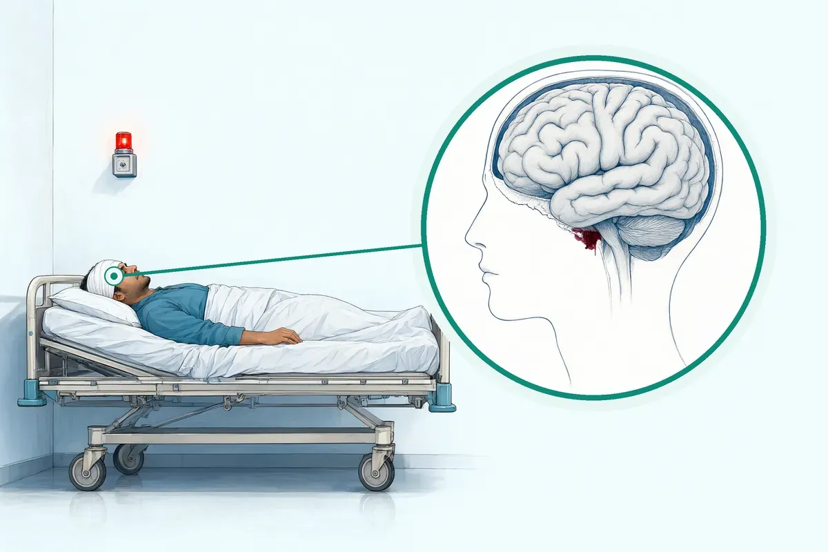
- Operation time
- 1 to 4 hours (emergency craniotomy/craniectomy)
- Anaesthesia
- General anaesthesia
- Recovery
- ICU stay days to weeks, neuro-rehabilitation often prolonged
- Price
- ₹1,50,000 to ₹5,00,000 (emergency surgery package)typical range in India; ask us for an estimate
Head injury disrupts everything
A sudden blow leaves you confused, unconscious, or unable to move
Bleeding or swelling inside the skull builds pressure fast. Every minute of delay can worsen the damage. You need a team that acts at once.
-
Loss of consciousness
May be brief or prolonged, signalling brain injury.
-
Rising intracranial pressure
Swelling or bleeding compresses vital brain structures.
-
Skull fracture risk
Bone fragments can tear the brain covering.
-
Long recovery road
Rehabilitation is often prolonged after the acute phase.
Axons in the white matter are especially vulnerable to mechanical loading during traumatic brain injury.
Why Jaipur Hospital
You are terrified for your loved one and overwhelmed by the speed of decisions and the unknown cost.
Your care plan
Your first steps with us
- 1 Call ambulance or arrive at emergency
- 2 Rapid CT scan and neurosurgical review
- 3 Emergency surgery if pressure is critical
- 4 ICU care then neuro-rehabilitation
How the operation is done
Under general anaesthesia, the surgeon opens the skull to relieve pressure, remove clots, or repair fractures. The bone flap may be kept out if swelling persists. Operation usually takes one to four hours.
-
Decompressive Craniectomy
Bone flap removed to allow brain swelling to expand without compression.
-
Evacuation of Haematoma
Craniotomy to remove acute subdural, epidural, or intracerebral clots.
-
Skull Fracture Elevation/Repair
Depressed bone fragments lifted and dural tears repaired.
-
ICP Monitoring & EVD
Invasive pressure sensor and ventricular drain placed for continuous monitoring.
Why patients trust us
- 1991 Serving Jaipur for 35 years
- NABH Accredited hospital
- 1 Neurosurgery and Neurology consultants
- 24×7 Emergency, ICU and ambulance
- 25 Cashless schemes and insurers
What research shows about this treatment
-
ICU stay median 9 to 12 days
Severe TBI patients in monitoring trials spent a median of 9 to 12 days in ICU.
-
6-month mortality 39 to 41%
Mortality at six months was 39% with pressure monitoring and 41% with clinical examination alone.
-
Decompressive craniectomy lowers mortality
Surgery for refractory pressure reduced death rates compared to medical care alone.
If it is left untreated
Untreated bleeding or swelling raises intracranial pressure, causing brain herniation, long-term disability, or death. Early surgery reduces pressure and limits secondary injury.
Life after treatment
Most people spend days to weeks in ICU, then move to ward and rehabilitation. Recovery continues for months with therapy.
-
ICU to ward transfer
Most people move from ICU to a general ward once stable.
-
Prolonged rehabilitation
Physiotherapy, speech and cognitive therapy often continue for months.
-
Gradual functional return
Many regain independence in daily activities over time.
-
Regular follow-up
Scheduled OPD reviews monitor healing and adjust therapy.
Frequently asked questions
Questions patients ask about head injury and neurotrauma
Which problems are assessed under head injury and neurotrauma?
In short
Jaipur Hospital assesses traumatic brain injury, skull fractures, and intracranial haemorrhage under its head injury and neurotrauma service. The 24x7 emergency team evaluates the severity of brain injury using clinical examination and imaging. Patients with raised intracranial pressure, haematomas, or compound depressed fractures are triaged for immediate intervention.
In detail
The assessment covers a spectrum from mild concussion to severe diffuse axonal injury. Evidence shows axons in white matter are especially vulnerable to mechanical loading during trauma. Clinical grading uses the Glasgow Coma Scale, which scores eye opening, verbal response, and motor response.
Lower scores correlate with higher likelihood of CT or MRI abnormalities and elevated blood biomarkers of glial and neuronal injury. Injuries are categorised as mild (GCS 13–15), moderate (GCS 9–12), or severe (GCS 3–8). This classification guides the urgency and type of investigation required.
A common belief
Many people think only loss of consciousness matters.
What actually happens
In reality, confusion, amnesia, or worsening headache after a fall can signal serious bleeding or swelling that needs immediate scanning.
Your next step at Jaipur Hospital
If you or your attendant suspect a head injury, proceed directly to the 24x7 emergency department at Jaipur Hospital. The neurosurgery team will perform an immediate clinical evaluation and arrange urgent CT scanning.
Based on findings, they will advise on observation, ICP monitoring, or emergency surgery. Bring any prior imaging or referral notes.
How will the doctor decide which tests I need for head injury?
In short
The neurosurgeon at Jaipur Hospital decides tests based on Glasgow Coma Scale score, mechanism of injury, clinical examination findings, and symptom progression. A CT head is standard for moderate to severe injury or any focal deficit. MRI may be added for suspected diffuse axonal injury or brainstem lesions. ICP monitoring is considered for severe TBI with refractory raised pressure.
In detail
Evidence supports a tiered approach. Lower GCS values correlate with higher likelihood of CT and MRI abnormalities. Blood biomarkers GFAP and UCH-L1 rise with injury severity. Intracranial pressure monitoring is standard of care for severe TBI.
In studies, care guided by ICP monitoring did not show superiority over imaging and clinical examination alone for functional outcomes at six months. The surgeon weighs radiation exposure, diagnostic yield, and urgency. For compound depressed fractures, CT with bone windows guides elevation planning. For suspected haematoma, non-contrast CT is the first-line investigation. The decision is individualised at the bedside.
A common belief
Some believe every head injury needs an MRI.
What actually happens
In acute trauma, CT is faster, better for bleeding and fractures, and guides emergency surgery. MRI is reserved for subtle axonal injury or when CT is inconclusive. ICP monitoring is not routine for all severe TBI.
Your next step at Jaipur Hospital
The doctor will explain which scans are needed and why. Most emergency imaging is done within the hospital's 24x7 radiology department. If ICP monitoring or EVD is advised, the procedure, risks, and ICU stay implications will be discussed with your attendant. Consent is obtained before any invasive monitoring.
How soon should I expect improvement after head injury surgery?
In short
Improvement after head injury surgery at Jaipur Hospital varies widely and cannot be predicted by a fixed timeline. The operation lasts one to four hours, but recovery in the ICU may last days to weeks. Consciousness, limb strength, and speech may change slowly, and neuro-rehabilitation is often prolonged.
In detail
Evidence from studies shows that axonal degeneration can continue for years after injury, and functional outcomes at three and six months differ greatly between patients.
Decompressive craniectomy lowers mortality compared to medical care alone, but it increases the rate of vegetative state while lowering severe disability. The Glasgow Outcome Scale at six months captures a wide spectrum from death to good recovery.
ICU stay in monitored groups averaged nine to twelve days in research settings. Your surgeon will track pupil response, motor scores, and infection markers daily to gauge progress.
A common belief
People often expect a quick bounce-back like after routine surgery.
What actually happens
Brain injury is different. Studies show recovery is measured at three and six months, and some changes continue for years. The first operation sets the stage, but the long road is rehabilitation.
Your next step at Jaipur Hospital
Expect daily assessments in the ICU and a step-down to the ward when stable. Physiotherapy, speech therapy, and occupational therapy start early and continue after discharge. An attendant should be trained for home care such as positioning, feeding, and medication timing.
Which changes in consciousness, headache, vomiting, or behaviour need emergency help after head injury?
In short
Go to the 24x7 emergency department at Jaipur Hospital immediately if the patient becomes difficult to wake, shows confusion that was not there before, vomits repeatedly, develops a severe or worsening headache, has a seizure, or shows weakness on one side of the body. Unequal pupil size or loss of consciousness are critical warning signs.
In detail
Evidence links lower Glasgow Coma Scale scores to higher likelihood of CT abnormalities and raised intracranial pressure. A drop of two or more points on the GCS triggers urgent imaging. Persistent vomiting suggests rising pressure or brainstem irritation.
New agitation, aggression, or apathy can indicate frontal lobe injury or expanding haematoma. The neurosurgical team uses pupil reaction, motor response, and verbal response to grade severity. Invasive ICP monitoring may be placed if the clinical picture is unclear but deterioration is suspected.
A common belief
Some think a mild headache or single vomit is fine.
What actually happens
But a change in consciousness or repeated vomiting often means pressure is rising inside the skull. The Glasgow Coma Scale is used by doctors to catch these changes early. At home, any drop in alertness is a red flag.
Your next step at Jaipur Hospital
Do not wait for a scheduled appointment. Call the ambulance at 0141-2551500 or +91-7414-001-001 or bring the patient directly to the emergency entrance. An attendant should stay with the patient and note the time symptoms started. The neurosurgery team is available round the clock.
How can I decide whether to seek advice about extradural haematoma evacuation?
In short
Extradural haematoma evacuation is an emergency neurosurgical procedure to remove blood between the skull and the dura mater. Jaipur Hospital provides 24x7 emergency management for this condition. You should seek immediate advice if a head injury is followed by a brief loss of consciousness, a lucid interval, and then rapid neurological deterioration such as worsening headache, vomiting, or weakness on one side of the body.
In detail
An extradural haematoma is usually caused by a skull fracture tearing an artery, most often the middle meningeal artery. Blood accumulates rapidly under pressure, compressing the brain. The standard treatment is an emergency craniotomy to evacuate the clot and stop the bleeding.
Evidence shows that early surgical evacuation, ideally within hours of symptom onset, is strongly associated with better functional outcomes and lower mortality. Delay increases the risk of irreversible brain injury from pressure and herniation.
The surgical team will assess the haematoma size, thickness, and midline shift on CT scan to decide on urgent surgery.
A common belief
20-45 s: Many people think if the patient wakes up they are safe.
What actually happens
That lucid interval is actually the warning window. Waiting at home risks long-term damage from rising pressure.
Your next step at Jaipur Hospital
If you or your attendant notice the classic pattern of deterioration after head trauma, call the Jaipur Hospital emergency line at 0141-2551500 or bring the patient directly to the 24x7 emergency department. Do not wait for symptoms to improve. The neurosurgery team is available round the clock for immediate CT scanning and operation theatre readiness.
What changes in strength, sensation, speech, vision, or bladder control need urgent assessment?
In short
Any new or worsening neurological deficit after head injury requires urgent assessment. Jaipur Hospital provides 24x7 emergency evaluation for these signs. Sudden weakness in an arm or leg, numbness, slurred speech, double vision, loss of vision, or inability to pass urine may indicate expanding haematoma, brain swelling, or brainstem compression needing immediate imaging and possible surgery.
In detail
Focal neurological signs localise the injury. Hemiparesis suggests contralateral cortical or subcortical compression. Cranial nerve palsies, especially third nerve dilatation, indicate uncal herniation. Speech arrest or aphasia points to dominant hemisphere involvement. Visual field defects signal occipital lobe or optic radiation injury.
Urinary retention or incontinence can reflect frontal lobe or spinal cord involvement. These deficits often progress rapidly as intracranial pressure rises. The Glasgow Coma Scale and pupil reactivity are monitored alongside focal signs. Early CT scan guides surgical decompression or haematoma evacuation to reverse or halt progression.
A common belief
20-45 s: People often hope the weakness will pass.
What actually happens
But these deficits usually mean a clot or swelling is pressing on vital brain areas. Delay can make the damage long-term.
Your next step at Jaipur Hospital
If you or your attendant notice any new neurological change after head trauma, call the Jaipur Hospital emergency number 0141-2551500 or come directly to the emergency department. Do not wait for the next OPD. The neurosurgery team will perform immediate CT and intervene if a surgical lesion is found.
Which symptoms should prompt me to see a specialist?
In short
You should see a neurosurgery specialist at Jaipur Hospital if you experience persistent headache, vomiting, confusion, memory loss, or drowsiness after a head injury. Any loss of consciousness, seizure, weakness in limbs, or clear fluid leaking from the nose or ears also warrants urgent specialist review. These symptoms may indicate raised intracranial pressure or structural brain injury.
In detail
Evidence indicates that depending on severity, traumatic brain injury can manifest acutely as immediate loss of consciousness or confusion and persist as coma or cognitive dysfunction. Lower Glasgow Coma Scale values are associated with loss of consciousness and longer post-traumatic amnesia.
Blood-based biomarkers such as GFAP and UCH-L1 rise with neuronal and glial injury, correlating with lower GCS scores. Progressive neurological decline, unequal pupils, or new focal deficits suggest expanding haematoma or worsening oedema.
Even seemingly mild symptoms like persistent headache or concentration difficulty can reflect diffuse axonal injury. Early specialist assessment allows timely imaging and intervention before irreversible damage occurs.
A common belief
People often dismiss a 'mild' knock to the head.
What actually happens
But diffuse axonal injury can cause ongoing cognitive problems without obvious signs on initial scan. Worsening headache or personality change days later can signal a slow bleed.
Your next step at Jaipur Hospital
Do not wait for symptoms to worsen. Attend the OPD or emergency department at Jaipur Hospital with an attendant. The neurosurgeon will take a detailed history, perform a neurological examination, and order CT or MRI as needed. If you have prior imaging, bring those films.
How will the results change my care plan for head injury?
In short
Test results directly determine whether you are managed with observation, medical therapy, ICP monitoring, or emergency surgery at Jaipur Hospital. A CT showing acute haematoma with mass effect typically leads to urgent craniotomy for evacuation. Diffuse injury with raised ICP may require decompressive craniectomy or EVD. Stable scans with mild symptoms allow monitored observation.
In detail
Evidence shows that imaging findings and ICP values guide specific interventions. Decompressive craniectomy for refractory ICP above 25 mm Hg reduced mortality but increased vegetative state rates compared to medical care alone.
Rates of severe disability were lower with surgery, while moderate disability and good recovery rates were similar. ICP monitoring did not prove superior to clinical and imaging-based management for six-month functional outcomes.
The neurosurgeon integrates scan findings, GCS trend, and ICP data to choose between craniotomy, craniectomy, haematoma evacuation, fracture elevation, or non-operative management. Each pathway carries different ICU duration and rehabilitation needs.
A common belief
A common belief is that surgery always gives a better outcome.
What actually happens
Studies show decompressive craniectomy lowers mortality but can increase vegetative state rates. The team balances survival against quality of life when recommending surgery versus continued medical care.
Your next step at Jaipur Hospital
After imaging or monitoring results are available, the surgeon will discuss the recommended pathway with you and your attendant. If surgery is advised, the typical package range is ₹1,50,000 to ₹5,00,000 with 7 to 21 nights hospital stay. ICU stay is usually days to weeks. Rehabilitation planning begins early.
What should I do if symptoms persist or worsen after head injury treatment?
In short
If symptoms persist or worsen after head injury treatment at Jaipur Hospital, contact the neurosurgical team or return to the 24x7 emergency department without delay. New or increasing headache, repeated vomiting, drowsiness, confusion, weakness, seizures, or unequal pupils may signal rising intracranial pressure, re-bleeding, or infection that needs urgent re-evaluation.
In detail
Evidence indicates that intracranial pressure can rise again after initial surgery, and haematomas can re-accumulate. The Glasgow Coma Scale is used repeatedly to detect deterioration; a drop of two or more points triggers an urgent CT scan.
Invasive ICP monitoring or external ventricular drainage may be in place to give real-time pressure data. Wound swelling, fever, or CSF leak from the nose or ear also need immediate review.
The first operation is critical, but complications can appear days later, so continuous monitoring in ICU and ward is standard.
A common belief
Patients sometimes think the danger is over once surgery is done.
What actually happens
In fact, re-bleeding, swelling, or infection can develop days later. The Glasgow Coma Scale is checked repeatedly in hospital for this reason. At home, any drop in alertness is an emergency.
Your next step at Jaipur Hospital
Keep the discharge summary and medication list handy. If the patient is at home, an attendant should watch for the warning signs above and call the hospital ambulance at 0141-2551500 or +91-7414-001-001. Do not wait for the next scheduled follow-up if there is any change.
Could symptoms worsen after an initially reassuring assessment for head injury?
In short
Yes, symptoms can worsen after an initially reassuring assessment for head injury. At Jaipur Hospital, patients are observed for this reason. A lucid interval can be followed by rapid deterioration due to expanding epidural or subdural haematoma, delayed brain swelling, or secondary injury cascades.
In detail
Evidence shows that axonal injury evolves over hours to days, and intracranial pressure can rise after a normal initial scan. Studies report that lower GCS values correlate with blood biomarkers of ongoing glial and neuronal injury.
The Glasgow Coma Scale is repeated at intervals because a patient who talks and obeys commands can suddenly decline. Observation protocols typically include half-hourly to hourly neurological checks in the emergency department and ICU. CT scan is repeated if the GCS drops or new signs appear.
A common belief
People often think a normal CT scan means everything is fine.
What actually happens
But pressure can rise hours later, and microscopic axonal injury continues. The Glasgow Coma Scale is checked repeatedly for this exact reason. A reassuring first check does not rule out later trouble.
Your next step at Jaipur Hospital
If you are sent home after observation, the discharge advice includes a list of warning signs for the attendant to watch. Return immediately if drowsiness increases, vomiting starts, headache worsens, or behaviour changes. Do not assume the danger has passed.
FAQ library
Questions and answers about head injury and neurotrauma
When you contact us
Get a neurosurgical opinion now
Contact Jaipur Hospital for an emergency assessment, scan review, and a clear plan with cost estimate and cashless help.
- A consultation with a neurosurgeon in the OPD
- Your scans and reports reviewed, with tests done in-house if you need them (laboratory, X-ray and imaging)
- Your options explained in plain words, including whether you need surgery at all
- A cost estimate before you decide
- Help with cashless treatment under RGHS, MAA Yojana, CGHS and empanelled insurers and TPAs
- Follow-up visits after your treatment
Overview
Head Injury and Neurotrauma: what to know
What is a Head Injury and Neurotrauma
A head injury happens when a sudden force hits the head and damages the brain, the skull, or the blood vessels inside the skull. The brain floats in fluid inside the hard skull. A hard blow can make the brain move back and forth, stretching or tearing the long nerve fibres called axons.
This tearing is known as diffuse axonal injury and it can happen even in mild injuries such as concussion. The damage may be only on the surface, like a scalp cut, or it may go deeper and cause bleeding inside the skull.
Bleeding can collect as a clot between the skull and the brain covering (epidural haematoma), under the brain covering (subdural haematoma), or inside the brain tissue itself (intracerebral haematoma). A skull fracture is a break in the bone that protects the brain.
When the bone is pushed inward it is called a depressed fracture and it can tear the brain covering. All of these problems can raise the pressure inside the skull. High pressure squeezes the brain and cuts off its blood supply, which can cause more damage within minutes to hours.
Symptoms and warning signs that need urgent care the same day
- Worsening headache
- Repeated vomiting
- Unequal pupils
- Weakness or numbness in a limb
- Slurred speech
- Seizures
Symptoms depend on how bad the injury is and which part of the brain is affected. A person may lose consciousness right away or feel confused and dazed. Some people do not pass out but cannot remember what happened just before or after the hit; this is called post-traumatic amnesia.
Other common signs are:
- Headache that gets worse
- Repeated vomiting
- One pupil larger than the other
- Weakness or numbness in an arm or leg
- Slurred speech
- Seizures
- Increasing drowsiness
In a baby or young child, watch for persistent crying, refusal to eat, or a bulging soft spot on the head. Any of these signs after a head knock mean you must go to the emergency department at once. At Jaipur Hospital the emergency team is ready 24×7 to assess and stabilise you without delay.
Causes and who is at higher risk
- Two-wheeler riders without helmets
- High risk in our city
- Older adults on blood thinners
- Minor falls can cause bleeds
- Young children
- Softer skulls, larger heads
The most common causes are:
- Road traffic crashes
- Falls from height or on level ground
- Assaults
- Sports injuries
Two-wheeler riders without helmets, pedestrians hit by vehicles, and construction workers are at high risk in our city. Older adults fall more often because of balance problems or weak bones, and even a minor fall can cause a bleed if they take blood-thinning medicines.
Young children have softer skulls and larger heads relative to their bodies, so a fall from a bed or stairs can cause serious injury. People who have had a previous head injury are more vulnerable to another one. Alcohol or drug use raises the chance of an accident and can mask the early symptoms.
How it is diagnosed: examination and tests
- Glasgow Coma Scale check Eye, verbal, motor scores
- CT scan of the head First and fastest test
- MRI if needed More detail later
- Pressure monitoring For severe injury
When you arrive, the doctor checks your level of consciousness using the Glasgow Coma Scale. The scale scores eye opening, verbal response, and motor response separately, for example E4 V4 M6. The three scores are often added to give a total from 3 to 15.
A total of 13 to 15 is called mild, 9 to 12 moderate, and 3 to 8 severe traumatic brain injury. A lower score means a higher chance of finding abnormalities on a CT or MRI scan. Blood tests for proteins such as GFAP and UCH-L1 are also higher when the injury is more severe.
A CT scan of the head is the first and fastest test to show bleeding, fractures, or brain swelling. MRI may be done later for more detail.
If the injury is severe, the team may place a small probe through the skull to measure the pressure inside the head continuously; this is called intracranial pressure monitoring and is considered the standard of care for severe traumatic brain injury. An external ventricular drain may be placed at the same time to drain excess fluid and lower the pressure.
Treatment options without surgery and when surgery is advised
Not every head injury needs an operation. Mild injuries with a normal CT scan and a good conscious level are often managed with close observation, pain control, and rest. The nursing team watches for any change in pupil size, limb strength, or alertness.
If the pressure inside the skull starts to rise, medicines such as mannitol or hypertonic saline may be given through a drip to shrink the brain slightly and buy time. Sedation and a breathing tube may be used to keep the patient still and lower the brain's oxygen demand.
Surgery is advised when there is a large blood clot pressing on the brain, a depressed skull fracture pushing on the brain covering, or when the pressure inside the skull stays high despite medical treatment. The neurosurgeon will explain which operation fits your situation.
Common procedures include evacuation of the haematoma through a craniotomy, elevation and repair of a compound depressed fracture, or a decompressive craniectomy where a bone flap is removed to give the swollen brain room to expand. Invasive pressure monitoring and external ventricular drainage may be done alone or with these operations. Your surgeon will discuss the plan with you and your attendant before proceeding.
Preparing for head injury and neurotrauma surgery
Do
- Tell the doctor about blood thinners
- Bring referral note and imaging
- Have an attendant stay with you
Head injury surgery is usually an emergency. You arrive through the 24×7 emergency department, where the team checks your level of consciousness, pupil reaction, and vital signs. A CT scan of the head is done immediately to show the type and size of any bleed or fracture. Blood tests include haemoglobin, coagulation profile, blood group, electrolytes, and kidney function.
If you take blood thinners or antiplatelet medicines, tell the doctor right away; these may need to be reversed before surgery. You will not be allowed to eat or drink for at least six hours before anaesthesia. If you are referred from another hospital, bring the referral note, previous imaging films or CDs, and a list of current medicines.
An attendant should stay with you to sign consent forms and coordinate with the team. The hospital provides a canteen with dietary meals and an in-house pharmacy for any urgent medicines.
How head injury and neurotrauma surgery is done
- Decompressive craniectomy
- Removes bone flap to relieve pressure
- Evacuation of haematoma
- Removes blood clots via craniotomy
- Compound depressed fracture repair
- Elevates bone fragments and repairs dura
The operation is performed under general anaesthesia and typically takes one to four hours. Your surgeon chooses the technique based on the CT findings and your clinical condition. Decompressive craniectomy removes a bone flap to relieve refractory raised intracranial pressure. Evacuation of haematoma uses a craniotomy to remove acute subdural, epidural, or intracerebral blood clots.
For a compound depressed skull fracture, the bone fragments are elevated and the dura is repaired. Invasive intracranial pressure monitoring or an external ventricular drain may be placed to measure and manage pressure inside the skull. The procedure is carried out in modular operation theatres with laminar airflow and HEPA filters. Intraoperative imaging such as a C-arm may be used.
After surgery you are shifted to the intensive care unit for continuous monitoring of neurological status, blood pressure, and intracranial pressure.
Risks and possible complications of head injury and neurotrauma surgery
- Bleeding
- May need a second surgery
- Infection
- Wound or meningitis, treated with antibiotics
- Seizures
- Anti-seizure medicines often given
- CSF leak
- Fluid leak from nose or wound
All brain surgery carries risks. General anaesthesia can cause breathing or heart problems. Bleeding may occur during or after the operation and may need a second surgery. Infection of the wound or the brain coverings (meningitis) is possible and is treated with antibiotics. Brain swelling can worsen after surgery and may require further decompression.
Seizures can develop; anti-seizure medicines are often given for a period. Stroke, weakness of limbs, speech difficulty, or cranial nerve injury can happen depending on the area operated on. A cerebrospinal fluid leak from the nose or wound may need repair.
In studies of decompressive craniectomy for refractory raised intracranial pressure, mortality at six months was lower with surgery than with medical care alone, but the rate of vegetative state was higher with surgery. Rates of severe disability were lower with surgery, while moderate disability and good recovery rates were similar between groups.
Serious adverse events were distributed similarly in monitored and non-monitored management strategies. Your surgeon will discuss the risks that apply to your specific injury.
Recovery and aftercare after head injury and neurotrauma surgery
- ICU stay Days to weeks
- Physiotherapy starts Early in ward
- Follow-up after discharge Outpatient or rehab centre
After surgery you stay in the intensive care unit for days to weeks. The team monitors your level of consciousness, pupil size, limb movement, blood pressure, and intracranial pressure if a monitor is in place. You may be on a ventilator for a period. Pain is managed with intravenous medicines. The surgical wound is inspected daily.
Once stable, you move to a ward bed. The total hospital stay is typically seven to twenty-one nights, including ICU and ward time. Physiotherapy starts early to prevent chest infection, blood clots, and joint stiffness. Occupational therapy and speech therapy are arranged if needed. You may need a urinary catheter and a nasogastric tube for feeding initially. Before discharge, the team reviews medicines, wound care, seizure precautions, and follow-up appointments.
Call the hospital or visit the emergency department if you notice:
- Increasing drowsiness
- New weakness
- Worsening headache
- Vomiting
- Fever
- Wound swelling or discharge
- Clear fluid leaking from the nose or ear
- A seizure
Neuro-rehabilitation is often prolonged and may continue as an outpatient or at a specialised centre.
Results of head injury and neurotrauma surgery
Outcomes depend on the severity of the initial injury, age, pupil reaction, and how quickly surgery is done. In a large trial of patients with severe traumatic brain injury and refractory raised intracranial pressure, decompressive craniectomy resulted in lower mortality than medical care at six months. However, surgery was associated with higher rates of vegetative state.
Rates of severe disability were lower with surgery, while moderate disability and good recovery rates were similar between the two groups. The Extended Glasgow Outcome Scale distribution differed between groups.
In another study comparing intracranial pressure monitoring with imaging and clinical examination alone, six-month mortality was 39 percent in the monitoring group and 41 percent in the clinical group, with no significant difference in the primary outcome. Median ICU stay was twelve days with monitoring and nine days without.
Axonal degeneration can continue for years after injury and has been linked to later neurodegenerative changes. These results come from groups of patients in studies; your individual outcome will be discussed by your treating team based on your specific situation.
Cost and insurance for head injury and neurotrauma surgery in Jaipur
The typical range for an emergency surgery package in India is ₹1,50,000 to ₹5,00,000. The final bill depends on the type of operation, length of ICU stay, need for ventilation, medicines, implants, and any complications. ICU stay usually costs ₹10,000 to ₹15,000 per day, while a ward room costs ₹6,000 to ₹8,000 per day.
The hospital accepts:
- RGHS
- MAA Yojana
- CGHS
- A panel of private insurers and TPAs
If you have an individual health insurance policy, surgery is usually covered after a waiting period of two years from policy inception. For employer-provided group policies, coverage often starts from day one. Check your policy for room rent capping and co-payment clauses to avoid unexpected expenses. The hospital's insurance desk can help with pre-authorisation and claim processing. Ask the admission team for an estimate based on your planned procedure and expected length of stay.