

Plastic Surgery · Jaipur Hospital
Reconstructive Microsurgery in Jaipur
Complex wounds need specialised repair. Jaipur Hospital offers assessment and a clear surgical plan.
- Serving Jaipur since 1991 (35 years)
- NABH accredited
- 24×7 emergency & ICU
- Cashless: RGHS, MAA Yojana, CGHS + 22 insurers
- Operation time
- 4 to 12+ hours
- Anaesthesia
- General anaesthesia
- Recovery
- Flap monitoring 5-7 daysrehab 3-6 months
- Price
- ₹2,00,000 to ₹8,00,000+typical range in India; ask us for an estimate
Wounds that will not heal
You have a wound or defect that standard surgery cannot close.
After cancer removal, trauma, or failed grafts, you may face open wounds, exposed bone, or loss of function in the mouth, jaw, or limbs. Simple dressings or skin grafts are not enough when blood supply or tissue bulk is missing.
-
Cancer surgery defects
Tumour removal leaves large gaps in the jaw, tongue, throat, or face that need vascularised tissue.
-
Trauma and crush injuries
Severe limb or facial injuries with lost skin, muscle, or bone need tissue transfer to heal.
-
Failed previous repairs
Wounds that did not heal after grafts or local flaps often need free tissue transfer.
-
Lymphoedema swelling
Chronic arm or leg swelling after cancer treatment may need supermicrosurgical lymphatic bypass.
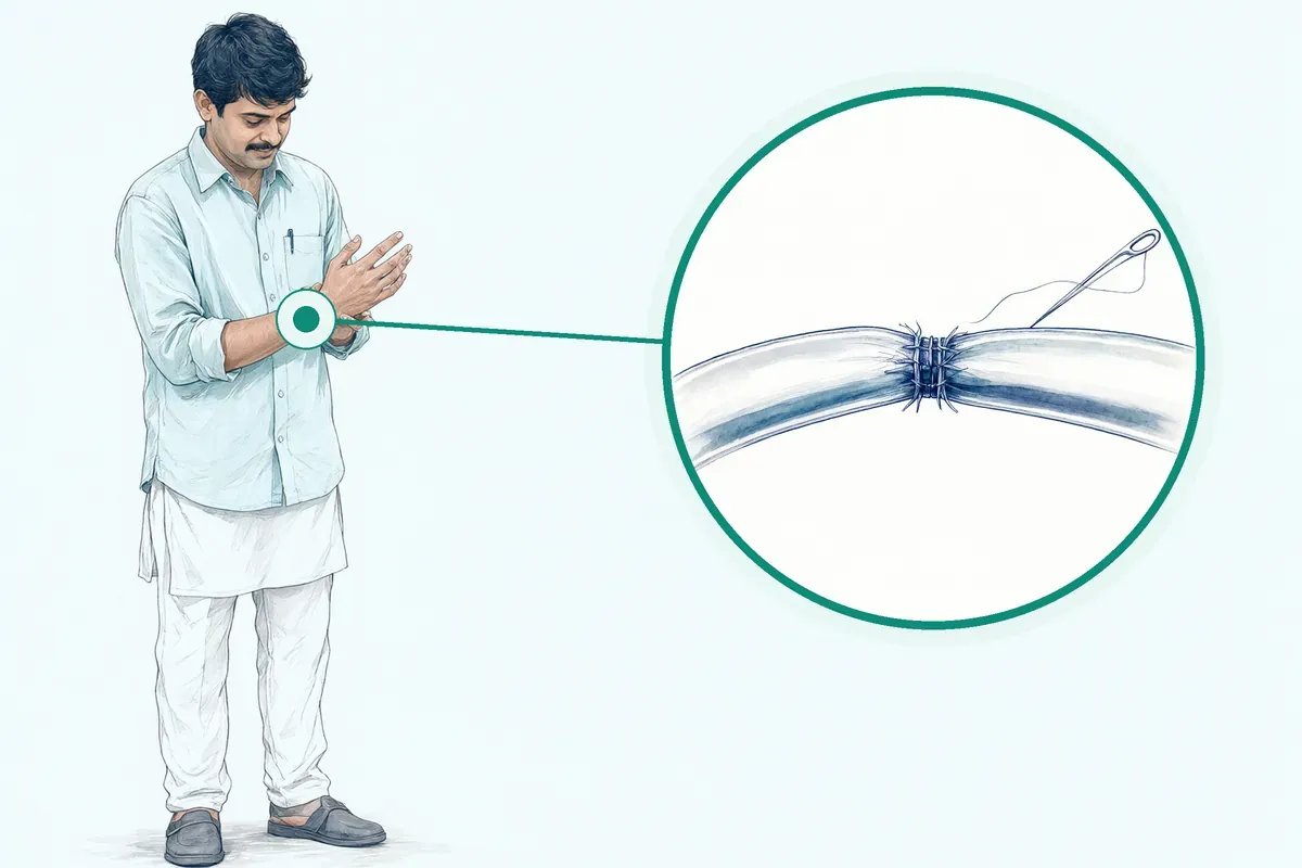
Free flap surgery transfers skin, muscle, or bone with its own blood supply to reconstruct defects from trauma or cancer.
Why Jaipur Hospital
You worry about losing the ability to speak, eat, or use your hand, and the long hospital stay that follows major surgery.
Your care plan
Your first steps with us
- 1 Call or visit OPD for surgical consultation
- 2 Complete in-house scans and pre-anaesthesia checks
- 3 Undergo microsurgical reconstruction with flap monitoring
- 4 Continue rehabilitation and follow-up after discharge
How the operation is done
Under general anaesthesia, the surgeon removes the tumour or clears the wound, harvests a vascularised flap from a donor site, and reconnects its tiny vessels to recipient vessels under a microscope. The flap is shaped and secured; the donor site is closed. Operation time is typically 4 to 12+ hours.
-
Free tissue transfer (free flaps)
Options include ALT, latissimus, fibula, or DIEP flaps for composite defects needing skin, muscle, or bone.
-
Digital or limb replantation
Arterial, venous, and nerve repair under microscope for amputated fingers or limb segments.
-
Lymphaticovenous anastomosis (LVA)
Supermicrosurgery connecting lymphatics to veins for lymphoedema reduction.
-
Perforator flap harvest
Muscle-sparing flaps based on reliable perforator anatomy for soft tissue coverage.
Why patients trust us
- 1991 Serving Jaipur for 35 years
- NABH Accredited hospital
- 1 Plastic Surgery consultants
- 24×7 Emergency, ICU and ambulance
- 25 Cashless schemes and insurers
What research shows about microsurgical reconstruction
-
about 7 days typical hospital stay
Most people stay in hospital for about seven days after free flap surgery if recovery is smooth.
-
re-exploration for flap congestion in about 3 in 100 cases
Operative re-exploration for flap congestion occurred in 2.7 percent of cases in a large series.
-
additional procedures needed in about 23 in 100 cases
Additional procedures occurred in 22.6 percent of cases after free tissue transfer.
If it is left untreated
Without reconstruction, complex wounds can lead to chronic infection, exposed bone or hardware, loss of limb function, difficulty speaking or swallowing, and progressive lymphoedema.
Life after treatment
Most people regain function and appearance over several months with rehabilitation.
-
Speech and swallowing
Most people recover close to preoperative speech and swallowing levels after one year.
-
Limb function
Physical therapy helps restore strength and mobility at the donor and recipient sites over months.
-
Lymphoedema relief
About 9 in 10 patients report subjective improvement after lymphaticovenous anastomosis.
-
Jaw reconstruction
Fibula flaps can support dental implants later to restore chewing ability.
Frequently asked questions
Questions patients ask about reconstructive microsurgery
Which problems are assessed under reconstructive microsurgery?
In short
At Jaipur Hospital, reconstructive microsurgery assesses complex tissue defects caused by cancer removal, severe trauma, burns, or chronic wounds that cannot heal on their own. The surgical team evaluates whether a free flap transfer, replantation, or lymphaticovenous anastomosis can restore form and function. Each case is reviewed for the size, depth, and location of the defect, the condition of nearby blood vessels, and your overall fitness for a long operation under general anaesthesia.
In detail
Reconstructive microsurgery is used when local tissue is not enough to cover a defect. Evidence shows free flaps bring their own blood supply, making them suitable for large or composite defects involving skin, muscle, bone, or nerve.
The team considers donor sites such as the forearm, thigh, leg, or back depending on the tissue needed. Microsurgical anastomosis under an operating microscope connects vessels as small as one millimetre. Lymphaticovenous anastomosis is assessed for lymphoedema when conservative measures fail. Your surgeon will match the flap type to the defect after imaging and clinical examination.
A common belief
Many people think a skin graft is enough for any wound.
What actually happens
Grafts rely on the wound bed for blood supply and fail on exposed bone, tendon, or radiated tissue. Flaps bring their own circulation, so they survive in difficult beds and can replace composite tissue like bone and muscle together.
Your next step at Jaipur Hospital
You will need a detailed clinical assessment and possibly imaging such as CT angiography to map vessels. The first consultation takes about 30 to 45 minutes.
If surgery is advised, pre-operative work-up includes blood tests, fitness review, and counselling about the 7 to 14 night hospital stay and 3 to 6 month rehabilitation.
How will the doctor decide which tests I need before reconstructive microsurgery?
In short
At Jaipur Hospital, the surgical team decides tests based on the defect location, planned flap type, and your medical history. Common tests include blood counts, coagulation profile, renal and liver function, ECG, chest X-ray, and imaging such as CT angiography or Doppler ultrasound to map recipient and donor vessels. Additional tests are ordered only if your history or examination suggests a specific risk.
In detail
Evidence shows pre-operative imaging of blood vessels is important to ensure a good blood supply for the transplant. CT angiography or MR angiography visualises the vessels at the recipient site and the chosen donor site. Doppler ultrasound is a bedside alternative.
Routine blood work assesses fitness for general anaesthesia and prolonged surgery. If you have diabetes, HbA1c is checked; if you have cardiac history, a cardiology opinion may be sought. The team follows enhanced recovery principles including pre-operative carbohydrate treatment, thromboprophylaxis, and antibiotic planning.
Tests are tailored — not every patient needs every investigation. The goal is to minimise surprises during the 4 to 12 hour operation and the 5 to 7 day flap monitoring period.
A common belief
Some patients expect a standard panel for everyone.
What actually happens
In microsurgery, vessel mapping is critical — a CT angiogram shows if the recipient vessels in your neck are usable and if the donor site vessels are adequate. Skipping this can lead to intraoperative surprises. Routine tests check you can tolerate 6 to 10 hours of anaesthesia safely.
Your next step at Jaipur Hospital
Tests are usually completed within 2 to 3 days before admission. The in-house diagnostic laboratory and imaging at Jaipur Hospital allow same-day reporting for most studies. Bring prior test results to avoid duplication. Insurance pre-authorisation for the procedure and tests is coordinated by the hospital TPA desk.
How soon should I expect improvement after reconstructive microsurgery?
In short
After reconstructive microsurgery at Jaipur Hospital, the first milestone is flap survival, which is monitored closely for five to seven days in hospital. You may stay seven to fourteen nights. Swelling and soreness improve over weeks, but complete healing and functional recovery typically take three to six months with rehabilitation.
In detail
In the first few days, the care team frequently checks the flap colour, temperature, and blood supply. Most people stay about seven days if recovery is smooth, though complex cases may need longer.
You may have a temporary feeding tube if the mouth or throat was reconstructed and possibly a tracheostomy for airway swelling. Speech, swallowing, and physical therapy start once you are stable.
Studies show significant decline in speech and swallowing early after tongue reconstruction, with most patients recovering close to preoperative level by one year. Outcomes depend on tumour size, resection area, reconstruction method, and adjuvant therapy.
A common belief
People often think the surgery is the finish line.
What actually happens
It is actually the start. The microscope work reconnects vessels, but your body must rebuild nerves, adapt muscles, and remodel scars. That takes months, not days. Rushing back to normal activity can stress the flap or donor site.
Your next step at Jaipur Hospital
Plan for a hospital stay of one to two weeks. Arrange for an attendant to help at home for at least two weeks after discharge. Follow-up visits will check flap healing, donor-site recovery, and therapy progress. You will receive specific timelines for returning to work or travel at your follow-up.
Why is a flap needed rather than a skin graft or a simpler closure?
In short
A flap is needed at Jaipur Hospital when the wound is too large, deep, or complex for a skin graft or direct closure. Flaps bring their own blood supply, so they survive on poorly vascularised beds, cover exposed bone or hardware, and replace composite tissue like bone, muscle, and skin together. Grafts rely on the wound bed for nutrition and cannot do this.
In detail
Flap surgery maintains its own blood supply, while skin grafting relies on the recipient site for blood supply. Grafts are not taken with their own blood supply. Local flaps keep their original supply but only reach adjacent defects.
Regional pedicled flaps rotate from nearby areas but have limited reach and bulk. Free flaps transfer tissue from distant sites with microvascular anastomosis, providing large volumes of skin, fat, muscle, or bone.
Flaps generally have longer healing time due to complexity but provide better cosmetic results and are ideal for larger defects or areas needing support. They are used when simpler options have failed or are unsuitable.
A common belief
Direct closure pulls edges together.
What actually happens
That works for small wounds. But stretching skin over a large gap cuts off its blood supply and causes necrosis. Pedicled flaps from nearby help, but they cannot reach everywhere or provide enough bulk. Free flaps solve reach, volume, and composite tissue needs in one stage.
Your next step at Jaipur Hospital
Your surgeon will explain why a graft or local closure will not work for your specific defect. Factors include wound size, depth, exposed structures, previous radiation, and infection. The decision is made after clinical examination and imaging.
How can I decide whether to seek advice about free flap reconstruction?
In short
You should seek advice about free flap reconstruction at Jaipur Hospital if you have a complex wound, a large tissue defect after cancer surgery, or trauma that cannot be closed simply. The procedure uses tissue from another part of your body with its own blood supply.
In detail
Free flap reconstruction is typically recommended for patients with extensive tissue loss due to trauma, surgical excision, or disease, and for those with chronic wounds or previous unsuccessful healing, according to the evidence.
It is often used after removal of head and neck cancers, including oral cavity, oropharyngeal, laryngeal, nasal, sinus, and salivary gland cancers, as well as advanced skin cancers. It is also used for composite defects needing bone, muscle, and skin.
The surgery is not suitable if you have poor overall health, such as uncontrolled diabetes, heart disease, or respiratory problems, because it relies on a healthy blood supply. Your surgeon will assess your condition and imaging to decide if you are a candidate.
A common belief
Some patients think a skin graft is enough for any wound.
What actually happens
But grafts rely on the wound bed for blood supply. Free flaps bring their own blood supply, making them better for large, deep, or radiated defects where grafts would fail. Your health must also support a long operation under general anaesthesia.
Your next step at Jaipur Hospital
If you have been told you need complex reconstruction, or if a wound is not healing, ask your doctor for a referral to Jaipur Hospital.
The surgical team will review your case, discuss donor site options, and explain the typical 7 to 14 night stay and 3 to 6 month rehabilitation.
How will appearance, function, and realistic expectations be discussed?
In short
At Jaipur Hospital, your surgeon will discuss appearance, function, and realistic expectations during the preoperative consultation. They use clinical photographs, diagrams, and sometimes 3D models to show typical outcomes. The conversation covers the donor site scar, the reconstructed area, and the likely timeline for recovery of speech, swallowing, or limb movement.
In detail
Evidence shows that outcomes are influenced by tumour size, area of resection, method of reconstruction, and adjuvant therapy. For tongue reconstruction, significant decline in speech and swallowing occurs early, but the majority recover close to preoperative level after one year.
Poorer outcomes follow resections involving both oral and base of tongue. Free flaps provide excellent long-term results and help regain function, appearance, and quality of life. Advances in microsurgical techniques continue to optimise functional and aesthetic results.
The surgeon will explain that scarring and sensation changes may last several months and that additional procedures such as dental implants may be needed later.
A common belief
Patients often focus only on the cancer removal.
What actually happens
The reconstruction plan is equally important. A flap that restores bulk helps speech and swallowing, but it takes months of therapy. The surgeon will be honest about scars, numbness, and the chance of further procedures.
Your next step at Jaipur Hospital
Ask for before-and-after photos of similar cases. Discuss your specific goals, such as returning to work or eating certain foods. The surgeon will outline the typical hospital stay of seven to fourteen nights and rehabilitation of three to six months.
Which symptoms should prompt me to see a specialist?
In short
See a reconstructive microsurgery specialist at Jaipur Hospital if you have a non-healing wound, exposed bone or tendon, a large defect after tumour removal, or persistent limb swelling despite compression therapy.
In detail
Symptoms that warrant referral include wounds that have not improved after several weeks of standard care, tissue loss exposing vital structures, defects after cancer resection where primary closure is not possible, and progressive lymphoedema with recurrent infections or skin changes.
In trauma, any amputation or crush injury with potential for replantation needs urgent evaluation. Chronic wounds with poor vascularity, radiation damage, or failed previous grafts also indicate microsurgical reconstruction. The surgeon assesses whether local, regional, or free flaps are needed based on defect size, location, and tissue requirements.
A common belief
Patients often hope dressings alone will heal deep defects.
What actually happens
Without vascularised tissue, these wounds can worsen, leading to infection, loss of function, or more extensive surgery later.
Your next step at Jaipur Hospital
Do not wait if the wound is enlarging, foul-smelling, or associated with fever. Ask your treating doctor for a referral or contact Jaipur Hospital directly. Early referral improves flap options and outcomes.
How will the results change my care plan for reconstructive microsurgery?
In short
Test results at Jaipur Hospital directly shape the surgical plan. If imaging shows unsuitable recipient vessels, the surgeon may choose a different flap or a vein graft. Abnormal blood counts or coagulation values may delay surgery until corrected. Poor cardiac or respiratory function may lead to a staged approach or non-surgical management. The care plan is adjusted to maximise flap survival and your safety.
In detail
Evidence highlights that flap success depends on meticulous planning, careful donor site selection, and precise microvascular technique. CT angiography defines vessel calibre, length, and quality at both recipient and donor sites.
If recipient vessels are damaged by prior surgery or radiation, the surgeon may use a vein graft from the leg or select a flap with a longer pedicle. Abnormal renal or liver function alters anaesthetic drug dosing and fluid management.
Uncontrolled diabetes impairs microvascular healing and increases infection risk; surgery may be postponed for optimisation. If cardiopulmonary reserve is limited, the team may split the procedure or consider a pedicled flap instead of a free flap.
The multidisciplinary team — surgeon, anaesthetist, physician — reviews all results together before finalising the plan. This reduces the chance of re-exploration for flap congestion, which occurs in about 2.7 percent of cases.
- This reduces the chance of re-exploration for flap congestion, which occurs in about 2.7 percent of cases.
A common belief
Patients sometimes think tests are just a formality.
What actually happens
In microsurgery, a CT angiogram showing a calcified recipient artery changes the whole operation — the surgeon may need a vein graft or a different flap with a longer pedicle. An HbA1c above 8 percent may postpone surgery for weeks. These decisions prevent flap failure and complications.
Your next step at Jaipur Hospital
You will discuss the results with your surgeon at a pre-operative visit. Any abnormal value will be explained with a clear action — correction, further test, or plan change. The typical timeline from completed tests to surgery is 1 to 2 weeks if all parameters are acceptable.
What should I do if symptoms persist or worsen after reconstructive microsurgery?
In short
If you notice increased pain, swelling, redness, bleeding, fever, or a change in the flap colour or temperature after discharge, contact Jaipur Hospital immediately. These signs may indicate infection, blood vessel blockage, or wound healing problems that need urgent assessment. Do not wait for a scheduled appointment if you are concerned.
In detail
Vigilant postoperative care is essential for monitoring complications such as blockages or clots in the newly joined vessels, infection, or failure of the incision to heal properly.
In studies, operative-site infection occurred in 4.7 percent of cases, lymphorrhea in 7.7 percent, and re-exploration for flap congestion in 2.7 percent. Additional procedures were needed in 22.6 percent of cases. Flap failure is rare but morbid and psychologically devastating when it occurs.
Early detection of vascular compromise allows surgical salvage. At home, watch the flap for pallor, darkness, coolness, or rapid swelling. Check donor sites for increasing pain, discharge, or separation.
- Additional procedures were needed in 22.6 percent of cases.
A common belief
Some patients wait to see if it improves, hoping it is just normal healing.
What actually happens
But a flap has a narrow window — usually hours — to fix a blood vessel problem. Waiting overnight can mean losing the reconstruction. It is always better to be checked and reassured than to wait too long.
Your next step at Jaipur Hospital
Keep the emergency numbers handy: 0141-2551500 and +91-7414-001-001. If you live far from Jaipur, go to the nearest hospital first and ask them to contact our team. Bring any discharge summary and medication list.
Where would donor tissue be taken from for reconstructive microsurgery, and what effects could that have?
In short
Common donor sites include the forearm, thigh, lower leg, back, abdomen and shoulder blade. The choice depends on the tissue type needed for the defect. Jaipur Hospital selects the donor site to match the reconstruction requirements while minimising functional impact. Each site has specific recovery considerations.
In detail
Evidence lists radial forearm flap for thin pliable tissue, fibula flap when bone is needed for jaw reconstruction, anterolateral thigh flap for large soft tissue volume, latissimus dorsi flap for scalp or large defects, rectus abdominis flap for muscle and skin, and scapula flap for bone with soft tissue.
Donor site morbidity includes temporary numbness, swelling, scarring and possible weakness. Physical therapy is often needed to regain strength and mobility at the donor site. A skin graft may be needed to close the donor site. The fibula flap can support dental implants for chewing restoration. ALT and latissimus flaps provide large soft tissue coverage.
A common belief
Patients often worry the donor site will be permanently disabled.
What actually happens
In reality, most people regain near-normal function with dedicated rehab. For example, the fibula flap removes a small calf bone but the leg remains stable, and the forearm flap leaves a visible scar but hand function returns with therapy.
Your next step at Jaipur Hospital
At the preoperative visit, the surgeon will explain the planned donor site, expected scar location, likely sensory changes and rehabilitation exercises. You will need to avoid heavy use of that limb for several weeks. Outpatient physiotherapy starts within 2 weeks of discharge.
FAQ library
Questions and answers about reconstructive microsurgery
When you contact us
Get a clear surgical plan
Contact Jaipur Hospital for an OPD consultation where a plastic surgeon reviews your reports, explains your options, and gives you a cost estimate before you decide.
- A consultation with a plastic surgeon in the OPD
- Your scans and reports reviewed, with tests done in-house if you need them (laboratory, X-ray and imaging)
- Your options explained in plain words, including whether you need surgery at all
- A cost estimate before you decide
- Help with cashless treatment under RGHS, MAA Yojana, CGHS and empanelled insurers and TPAs
- Follow-up visits after your treatment
Overview
Reconstructive Microsurgery: what to know
What reconstructive microsurgery is and how it works
Free flap
- Brings its own artery and vein
Skin graft
- No blood supply of its own
Reconstructive microsurgery is a specialised operation that moves tissue from one part of your body to another to repair a complex wound or defect. The surgeon uses an operating microscope to join tiny blood vessels and nerves, usually smaller than two millimetres, so the transferred tissue survives in its new location.
This is called a free flap transfer because the tissue is completely separated from its original blood supply and reconnected at the recipient site. The tissue can include skin, fat, muscle, bone or a combination, depending on what the defect needs.
Common donor sites are:
- Forearm
- Thigh
- Lower leg
- Back or abdomen
The choice depends on the type and amount of tissue required, the blood vessel anatomy, and your overall health.
A free flap is different from a skin graft. A graft has no blood supply of its own and relies on the wound bed to grow new vessels. A flap brings its own artery and vein, which the surgeon stitches to vessels near the defect under the microscope.
This makes flaps suitable for larger defects, areas with poor blood supply, or places that need bulk and structural support such as the jaw or tongue.
The operation typically takes four to twelve hours or more under general anaesthesia. Your surgical team plans the donor site and the recipient site together, often using imaging to check the blood vessels before surgery.
Symptoms and warning signs that need urgent care
- Drop in temperature
- Rapid swelling increase
- Bleeding from edges
Reconstructive microsurgery is not a treatment for a symptom you notice at home; it is a planned operation after cancer removal, major trauma, or when a chronic wound will not heal. However, after the surgery you must watch for signs that the flap is in trouble.
A change in the flap colour to pale or dusky purple, a drop in temperature, swelling that increases quickly, or bleeding from the edges can mean the blood vessels have clotted or the flap is congested.
These are emergencies that need the surgical team to re-explore the flap immediately, usually within the first five to seven days while you are still in hospital.
If you are at home after discharge and notice sudden swelling, redness spreading from the wound, fever, or the flap feels cold, contact the hospital at once. Early return to the operating theatre can save the flap.
Causes and who is at higher risk for needing reconstructive microsurgery
- Head and neck cancer removal
- Oral cavity, throat, voice box, sinuses, salivary glands, skin cancers
- Severe trauma
- Destroys skin, muscle and bone
- Chronic non-healing wounds
- After radiation, infection or previous surgery
- Large benign tumours
- Invade nearby structures needing wide excision
The most common reason for free flap reconstruction is removal of a head and neck cancer.
This includes cancers of the:
- Oral cavity (lip, tongue, gum, floor of mouth, hard palate)
- Oropharynx (soft palate, tonsils, base of tongue)
- Larynx (voice box)
- Nasal cavity and paranasal sinuses
- Salivary glands
- Advanced skin cancers of the head and neck
Other causes are severe trauma that destroys skin, muscle and bone; chronic wounds that fail to heal after radiation, infection or previous surgery; and benign tumours that are large or invade nearby structures and need wide excision.
Not everyone is a candidate for free flap surgery. You need a reasonably healthy blood vessel system for the microsurgical connections to work. Uncontrolled diabetes, significant heart or lung disease, and severe peripheral vascular disease increase the risk of flap failure.
Your surgical team will assess your overall health, smoking status, and the condition of the potential donor and recipient vessels before recommending this operation.
How the need for reconstructive microsurgery is diagnosed
The decision for free flap reconstruction is made by a multidisciplinary team after the primary disease is diagnosed and staged.
For cancer, this involves:
- A biopsy to confirm the tumour type
- Imaging such as CT, MRI or PET-CT to define the extent of the tumour and whether it involves bone or deep structures
- Examination under anaesthesia to assess the exact margins
For trauma or chronic wounds, the diagnosis is based on clinical examination of the defect size, depth, exposed structures (bone, tendon, nerve, vessel), and the quality of the surrounding tissue. The surgeon checks whether a simpler option like a local flap, regional pedicled flap, or skin graft can close the defect.
If the defect is too large, involves composite tissue loss, or the local tissue is damaged by radiation or scarring, a free flap becomes the preferred option.
Preoperative imaging of the donor site vessels (CT angiography or Doppler ultrasound) may be done to confirm anatomy and rule out vascular disease, especially if you have diabetes or a history of smoking.
Non-surgical options and when reconstructive microsurgery is advised
Skin graft
- Shallow wound with healthy bed
Local flap
- Tissue next to the defect
Regional pedicled flap
- Nearby area on intact vessel
Free flap
- Large, composite, or radiated defects
Before recommending free flap surgery, the team considers simpler methods. A skin graft may be enough for a shallow wound with a healthy bed. A local flap uses tissue next to the defect and keeps its original blood supply; it is useful for smaller defects.
A regional pedicled flap rotates tissue from a nearby area (for example, chest muscle to the neck) on its intact blood vessel pedicle; it avoids microsurgery but has limited reach and bulk.
Free flap reconstruction is advised when the defect is large, involves multiple tissue types (skin, muscle, bone), the recipient bed has poor blood supply from radiation or scarring, or when function such as speech, swallowing, or jaw continuity must be restored. It is also used for lymphaticovenous anastomosis (supermicrosurgery) to treat lymphoedema when conservative therapy has failed.
Your surgeon will explain the options that fit your specific defect, health status, and goals. The final plan is made jointly with you and your attendants after discussing the operation time, hospital stay of seven to fourteen nights, flap monitoring for five to seven days, and a rehabilitation period of three to six months.
The typical cost range in India is ₹2,00,000 to ₹8,00,000 or more, varying with the flap type, complexity, and any additional procedures.
Preparing for reconstructive microsurgery
Do
- Comfortable loose front-opening clothing
- Regular medicines in original strips
- Imaging films or reports
- Insurance or scheme cards
- Phone charger
Before your operation, your surgical team will assess your overall health and the specific defect that needs reconstruction. This assessment helps the team choose the most suitable donor site for the free flap, which may be the forearm, thigh, leg, or back depending on the type and amount of tissue required.
You may need imaging tests such as a CT angiogram or Doppler ultrasound to check the blood vessels at the donor site and the recipient area, ensuring a reliable blood supply for the transplant.
You will meet the anaesthesia team for a pre-anaesthesia check-up.
Tell them about every medicine you take, including:
- Blood thinners
- Diabetes tablets
- Insulin
- Herbal products
- Any supplements
Your surgeon and anaesthetist will advise which medicines to continue, pause, or adjust before surgery. If you have diabetes, heart disease, or a lung condition, good control of these problems before the operation lowers the chance of complications.
You will be asked to fast for at least six hours before general anaesthesia — no solid food, and usually clear fluids are allowed up to two hours before. The hospital will confirm the exact fasting time the day before surgery.
Pack a small bag for a stay of one to two weeks. Bring comfortable loose clothing that opens at the front, your regular medicines in their original strips, any imaging films or reports, your insurance or scheme cards (RGHS, MAA Yojana, CGHS, or private TPA details), and a phone charger. An attendant should plan to stay with you during the admission.
How reconstructive microsurgery is done
Reconstructive microsurgery uses an operating microscope to join very small blood vessels and nerves.
The main techniques available at Jaipur Hospital include:
- Free tissue transfer (free flaps) such as ALT, latissimus dorsi, fibula, and DIEP flaps for composite defects
- Digital or limb replantation with arterial, venous, and nerve repair under the microscope
- Lymphaticovenous anastomosis (LVA) for lymphoedema
- Perforator flap harvest for muscle-sparing reconstruction
Your surgeon will advise which technique suits your defect, the tissue needed, and your anatomy.
On the day of surgery, you are taken to the modular operation theatre and given general anaesthesia. The procedure typically lasts four to twelve hours or more, depending on the complexity.
The main steps are:
- Tumour or damaged tissue removal: The surgeon removes the cancer, injured tissue, or non-healing wound while preserving as much healthy structure as possible.
- Flap harvest: The chosen donor tissue — skin, fat, muscle, bone, or a combination — is carefully dissected with its tiny blood vessels (artery and vein) and sometimes a nerve.
- Recipient site preparation: The area that will receive the flap is cleaned and healthy blood vessels are identified in the neck or face for connection.
- Microvascular anastomosis: Under the microscope, the surgeon stitches the flap's artery and vein to the recipient vessels. This step, called anastomosis, restores blood flow to the transplanted tissue.
- Flap inset and fixation: Once blood flow is confirmed, the flap is shaped and sutured into place. If bone is transferred (for example, fibula for jaw reconstruction), small plates and screws may secure it.
- Donor site closure: The area where the flap was taken is closed directly or covered with a skin graft if needed.
After the operation, you are moved to the intensive care unit or a high-dependency ward for continuous flap monitoring.
Risks and possible complications of reconstructive microsurgery
- 4.7 percent Operative-site infection
- 7.7 percent Lymphorrhea
- 2.7 percent Re-exploration for flap congestion
- 22.6 percent Additional procedures needed
Free flap surgery is a major operation. While most flaps survive, complications can occur. In studies of free flap reconstruction, operative-site infection was reported in about 4.7 percent of cases. Lymphorrhea (leak of lymphatic fluid) occurred in about 7.7 percent of cases.
Re-exploration of the operation site for flap congestion — a blockage in the vein draining the flap — was needed in about 2.7 percent of cases. Additional procedures of any kind were required in about 22.6 percent of patients during the same admission.
Flap failure — complete loss of the transplanted tissue — is relatively rare but when it happens it is very morbid and psychologically devastating. Complications after free tissue transfer to the head and neck remain common.
Other risks include:
- Blockage or clots in the newly joined vessels
- Infection
- Wound healing problems at the flap or donor site
General anaesthesia carries its own risks such as heart or lung events, deep vein thrombosis, and reactions to medicines. Patients with uncontrolled diabetes, significant heart disease, or severe respiratory problems have a higher chance of complications and may not be suitable candidates for flap surgery.
Your surgical team will discuss the risks that apply to your specific operation and health status before you sign the consent form.
Recovery and aftercare after reconstructive microsurgery
After surgery you will stay in the hospital for flap monitoring, usually five to seven days, and the total hospital stay is typically seven to fourteen nights. In the first few days, nurses check the flap's colour, temperature, capillary refill, and Doppler signal every hour or two. You may have drains, bandages, or a splint to protect the reconstruction.
A temporary tracheostomy (breathing tube in the neck) is common if the reconstruction involves the tongue, throat, or jaw. It is usually removed once swelling settles and you can breathe safely. The stoma heals within a few days to weeks.
Many patients need a feeding tube — either a nasogastric tube through the nose for short-term use, or a gastrostomy tube through the abdomen if longer support is expected. The team will assess your swallowing and help you transition back to eating when it is safe.
Pain is managed with regular medication. Swelling and bruising are normal and improve over weeks. You may have temporary numbness or tightness at both the surgical site and the donor site.
Before discharge, the team will give you instructions on wound care, drain care if a drain goes home with you, medicines, and follow-up dates. You will need an attendant to help at home for the first one to two weeks.
In the first weeks at home:
- Keep the flap and donor site clean and dry as instructed.
- Avoid pressure, stretching, or trauma to the reconstructed area.
- Attend all follow-up appointments for wound checks and flap assessment.
- Start gentle movement as advised; if the flap came from your arm, leg, or thigh, you may need physiotherapy to regain strength and mobility.
- If the surgery affects speech or swallowing, a speech therapist will guide exercises.
Complete healing can take several months. Most people regain function and appearance, but some may need additional procedures such as dental implants if the jaw was reconstructed.
Call the hospital or visit the emergency department if you notice:
- Sudden colour change of the flap (pale, dusky, or very dark)
- Loss of warmth
- Rapid swelling
- Bleeding that soaks the dressing
- Fever above 38°C
- Increasing pain not relieved by prescribed medicines
- Any new breathing or swallowing difficulty
Results of reconstructive microsurgery
Free flap reconstruction is an indispensable option for complex defects of the head and neck, limbs, and other areas. In studies, these reconstructions provide excellent long-term results and help patients regain function, appearance, and quality of life after cancer surgery or trauma.
Functional recovery depends on the site and extent of resection. For patients with reconstruction limited to the oral tongue or base of tongue, significant decline in speech and swallowing is common in the early postoperative phase, but the majority recover close to preoperative level after one year.
When both the oral tongue and base of tongue are resected, speech and swallowing outcomes are poorer regardless of the flap type used. Overall results are influenced by tumour size, area of resection, method of reconstruction, and whether adjuvant radiotherapy or chemotherapy is given.
Specific flap types have recognised advantages:
- The radial forearm free flap is thin and pliable, making it suitable for restoring speech and swallowing after smaller cancers
- The fibula free flap provides bone strong enough to support dental implants, helping restore chewing ability
- The ALT and latissimus dorsi flaps can supply large amounts of soft tissue for extensive defects
For lymphoedema, supermicrosurgical lymphaticovenous anastomosis has shown reduction in excess limb circumference by about 49 percent and excess volume by about 57 percent in reported series. About 91 percent of patients reported subjective improvement, and approximately 65 percent were able to stop using compression garments at follow-up. However, about 12 percent of patients had no measurable improvement.
Advances in microsurgical technique and perioperative care continue to improve functional and aesthetic outcomes. Your surgeon will discuss the expected functional goals for your particular case.
Cost and insurance for reconstructive microsurgery in Jaipur
The typical cost range for reconstructive microsurgery in India is ₹2,00,000 to ₹8,00,000 or more. The final amount depends on the complexity of the defect, the type of flap used, whether bone or multiple tissue components are transferred, the duration of the operation, the length of ICU and ward stay, and any additional procedures or complications that arise.
Jaipur Hospital is a NABH-accredited multispecialty hospital established in 1991.
The hospital accepts patients under:
- RGHS
- MAA Yojana
- CGHS
- A panel of private insurers and TPAs
If you are covered under any of these schemes, bring your scheme card and any pre-authorisation documents at admission. The hospital's insurance coordination team can assist with the paperwork required by your scheme or insurer.
For patients paying out of pocket, the hospital will provide an estimate after the surgical plan is finalised. The estimate covers the operation, anaesthesia, implants if needed, ICU and ward stay, medicines, investigations, and standard nursing care. Items not included are typically special drugs, additional procedures, or extended stay beyond the planned period.
Discuss the estimate with the admission office before surgery so you understand what is covered and what may be extra.