

Plastic Surgery · Jaipur Hospital
Wound Care and Limb Salvage in Jaipur
When an open wound does not heal, our surgical team offers clinical evaluation, diagnostic imaging, and a structured treatment plan.
- Serving Jaipur since 1991 (35 years)
- NABH accredited
- 24×7 emergency & ICU
- Cashless: RGHS, MAA Yojana, CGHS + 22 insurers
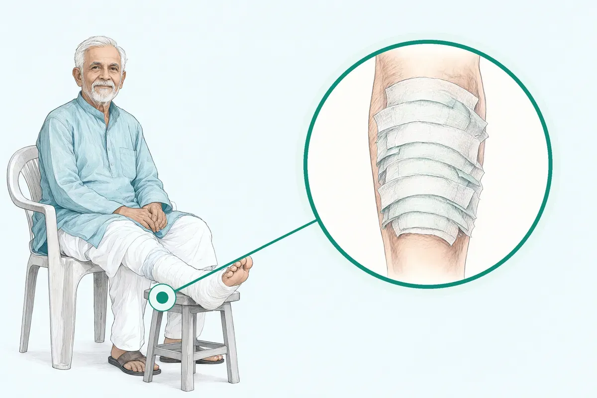
- Procedure time
- 1 to 4 hours per session
- Anaesthesia
- Local or Regional anaesthesia
- Recovery
- Daily dressings for 2 to 6 weeksphysio as needed
- Price
- ₹15,000 to ₹3,50,000typical range in India; ask us for an estimate
Wounds that refuse to heal
You are worried about losing your foot or leg
A chronic sore changes daily life. Frequent dressing changes take hours, pain limits movement, and uncertainty about limb amputation causes constant anxiety for you and your attendant.
-
Persistent open ulcers
Sores on the foot or leg stay open for weeks despite regular antiseptic dressings at home.
-
Loss of working days
Resting the affected foot keeps you away from work and lowers family earnings over time.
-
Dressing and travel expenses
Frequent visits to the chemist and clinics lead to rising out-of-pocket costs every month.
-
Fear of limb loss
Deep infections or blackening tissue raise concerns about whether the limb can be saved.
Medications, ulcer dressing, and periodic debridement accounted for 79.26% of direct medical costs in diabetic foot ulcer care.
Why Jaipur Hospital
You may worry about daily dressing costs, missing work, and whether an open wound will ever close.
Your care plan
Your first steps with us
- 1 Book an initial consultation at our OPD
- 2 Undergo diagnostic tests and tissue assessment
- 3 Review surgical options with your surgeon
- 4 Follow structured dressings and guided recovery
How surgical wound management works
Your surgeon cleans unhealthy tissue and prepares the wound bed. Procedures typically take 1 to 4 hours per session under local or regional anaesthesia, with hospital stay ranging from daycare to 5 nights.
-
Negative Pressure Wound Therapy (NPWT)
Your surgeon will discuss vacuum-assisted dressings to remove wound fluid and support granulation tissue.
-
Skin Grafting (Split/Full thickness)
Your surgeon will discuss transferring thin sheets of healthy skin to cover clean raw areas.
-
Local / Regional Flap Coverage
Your surgeon will discuss shifting nearby tissue with its blood supply over exposed bone or tendon.
-
Free Flap Reconstruction
Your surgeon will discuss microvascular transfer of tissue from another area for complex limb defects.
Why patients trust us
- 1991 Serving Jaipur for 35 years
- NABH Accredited hospital
- 1 Plastic Surgery consultants
- 24×7 Emergency, ICU and ambulance
- 25 Cashless schemes and insurers
Clinical findings in wound and limb salvage research
-
1.9 times
The relative risk of developing an infection was 1.9 times higher in control patients than in patients treated with NPWT.
-
52.55%
Study participants spent an average of 52.55% of their annual family income managing diabetic foot ulcers and complications.
-
1 to 2 weeks
Antibiotic therapy of 1 to 2 weeks often proves adequate for soft tissue infections, though severe disease may need longer.
If it is left untreated
Postoperative wound complications may lead to increased healthcare costs due to prolonged inpatient stay, repeat surgery, and the need for increased follow-up. Delayed care of progressive lesions and deep tissue breakdown can increase the risk of above-the-ankle amputation.
Expectations after treatment
A planned approach supports wound recovery through systematic care and guided rehabilitation.
-
Protected wound bed
Most people receive daily dressings for 2 to 6 weeks to protect newly formed skin.
-
Gradual mobility
Patients typically resume gentle walking with offloading footwear and physio as needed.
-
Routine clinic reviews
Regular OPD visits allow the surgical team to observe graft take and tissue stability.
Frequently asked questions
Questions patients ask about wound care and limb salvage
Which problems are assessed under wound care and limb salvage?
In short
Jaipur Hospital assesses chronic non-healing wounds, diabetic foot ulcers, venous leg ulcers, arterial ulcers, pressure injuries, infected wounds, and wounds with exposed bone or tendon. The service also evaluates limbs at risk of amputation due to severe infection, ischaemia, or complex soft tissue defects requiring reconstruction.
In detail
The assessment covers the wound bed, surrounding skin, vascular status, infection signs, and underlying causes such as diabetes, peripheral arterial disease, or neuropathy. Evidence shows that if non-infected ulcers do not heal after four to six weeks of optimal care, adjunctive treatments should be considered.
Vascular thresholds guide urgency: ankle pressure below 50 mmHg or ABI less than 0.4 warrants urgent imaging and revascularisation. The team also classifies the type and extent of tissue loss to match the appropriate reconstructive technique, from skin grafts to free flaps.
A common belief
Many people think only diabetic ulcers qualify, but venous ulcers, arterial ulcers, traumatic wounds, and post-surgical wounds that fail to heal are also assessed.
What actually happens
The key is whether the wound is stuck in the healing cascade or the limb is threatened.
Your next step at Jaipur Hospital
You will attend the OPD for a detailed examination, vascular assessment, and imaging if needed. The surgeon then outlines a plan that may include debridement, offloading, dressings, or surgery. Daily dressings typically continue for two to six weeks.
How will the doctor decide which tests I need?
In short
The surgeon at Jaipur Hospital selects tests based on wound type, duration, clinical signs of infection or ischaemia, and your medical history. Common tests include blood counts, inflammatory markers, blood sugar, HbA1c, renal function, wound swab for culture, ankle-brachial index, toe pressure, transcutaneous oxygen measurement, and imaging such as X-ray, Doppler ultrasound, CT angiography, or MRI.
In detail
Evidence guides the work-up: vascular thresholds (ankle pressure <50 mmHg, ABI <0.4, toe pressure <30 mmHg, TcPO2 <25 mmHg) trigger urgent vascular imaging and revascularisation consideration. Infection signs direct wound swabs and inflammatory markers. Neuropathic ulcers prompt offloading assessment and imaging for osteomyelitis.
The surgeon also reviews plain radiographs, CT, or MRI to define bony architecture and soft tissue extent before reconstructive planning. Each test answers a specific question: is there deep infection, is perfusion adequate for healing, is bone involved, and what reconstructive options are feasible.
A common belief
Patients sometimes expect every test at once.
What actually happens
But each test answers a specific question: is there deep infection, is blood flow enough for healing, is bone infected, what reconstruction is possible. The surgeon orders only what changes the plan, avoiding unnecessary cost and delay.
Your next step at Jaipur Hospital
Tests are done at the in-house diagnostic laboratory and radiology. Results are usually available the same day or next day. The surgeon reviews them with you and adjusts the plan — adding antibiotics, scheduling debridement, referring for revascularisation, or planning flap coverage.
How soon should I expect improvement after wound care and limb salvage treatment?
In short
Improvement after wound care and limb salvage treatment at Jaipur Hospital varies by wound type and technique used. Many patients see granulation tissue form within the first two to three weeks of Negative Pressure Wound Therapy or after debridement. Skin grafts and flaps take longer to mature, and full healing may need several weeks of daily dressings and physiotherapy.
In detail
Evidence shows that Negative Pressure Wound Therapy promotes angiogenesis and granulation tissue formation through mechanical strain and growth factor modulation, with studies reporting reduced infection and seroma rates compared with standard dressings.
In a controlled study, patients receiving early debridement, NPWT, revascularisation and skin graft or flap had significantly better healing rates, lower recurrence and lower amputation rates than those on standard care alone. Revascularisation improved ankle-brachial index and transcutaneous oxygen pressure significantly.
However, wound size may temporarily increase after initial debridement, and donor sites from grafts or flaps create secondary wounds that need care. Healing speed depends on blood supply, infection control, diabetes control, nutrition and adherence to offloading.
A common belief
Some people expect the wound to shrink immediately.
What actually happens
In reality, the wound often looks larger after the first debridement because dead tissue is removed. Healing is a stepwise process, not a straight line.
Your next step at Jaipur Hospital
Your surgeon will set review dates, usually weekly at first, to assess granulation, exudate and signs of infection. Dressings continue daily for two to six weeks as per protocol. Physiotherapy starts early to maintain joint mobility. Plan for time off work if the wound is on a weight-bearing area.
How will blood flow be assessed before wound care and limb salvage treatment?
In short
Before wound care and limb salvage treatment at Jaipur Hospital, blood flow is assessed using ankle-brachial index, toe pressure and transcutaneous oxygen pressure measurements. If ankle pressure is below 50 mmHg, ankle-brachial index is less than 0.4, toe pressure is below 30 mmHg, or transcutaneous oxygen pressure is less than 25 mmHg, urgent vascular imaging and revascularisation are considered. These thresholds guide the need for angiography or duplex ultrasound.
In detail
Guidelines recommend revascularisation when optimal treatment does not result in ulcer healing signs within four to six weeks, regardless of vascular test results. If an above-ankle amputation is being contemplated, revascularisation should first be considered as an option.
Individual factors such as the morphologic distribution of peripheral artery disease, autogenous vein availability, patient comorbidities and local operator expertise determine the revascularisation technique. Indications for vascular surgery include intractable rest pain, intractable foot ulcers, and impending or existing gangrene.
Intermittent claudication alone infrequently warrants bypass surgery. Pharmacologic treatments have not been proven to benefit perfusion. After revascularisation, perfusion is objectively measured to assess effectiveness; studies show significant improvement in ankle-brachial index and transcutaneous oxygen pressure post-revascularisation.
A common belief
Some patients think a pulse check is enough.
What actually happens
Objective pressure measurements are needed because pulses can be misleading, especially in diabetes where vessels are calcified.
Your next step at Jaipur Hospital
Your surgeon will order non-invasive vascular studies at the first visit. If results suggest significant ischaemia, angiography or CT angiography will be arranged. Revascularisation, if needed, is done before or with definitive wound coverage.
How can I decide whether to seek advice about complex wound flap reconstruction?
In short
Consider seeking advice when a wound exposes bone, tendon, or joint, when standard dressings and grafts have failed, or when the defect is too large for a skin graft alone. At Jaipur Hospital, the plastic surgery team evaluates each case to determine if a local, regional, or free flap is appropriate.
In detail
Flap reconstruction brings healthy, vascularised tissue to cover complex defects that cannot heal by secondary intention or skin grafting. Evidence indicates that flaps are indicated for exposed vital structures, large soft‑tissue loss, or irradiated beds.
The choice depends on defect size, location, donor‑site availability, and your overall health. Local and regional flaps use nearby tissue on a pedicle; free flaps transfer distant tissue with microvascular anastomosis. Both require careful preoperative imaging, optimisation of medical comorbidities, and postoperative monitoring.
The team discusses the expected functional outcome, donor‑site morbidity, and the likely number of procedures so you can make an informed decision.
A common belief
Patients often wait too long, hoping dressings will work.
What actually happens
Evidence shows that early flap coverage of exposed structures reduces infection risk, shortens hospital stay, and improves functional recovery compared with prolonged conservative care.
Your next step at Jaipur Hospital
If your wound shows exposed bone, tendon, or hardware, or if previous grafts have not taken, request a flap assessment at Jaipur Hospital. The surgeon will review imaging, check vascular status, and explain the options, risks, and recovery timeline.
How will appearance, function, and realistic expectations be discussed?
In short
At Jaipur Hospital, the surgical team holds a detailed preoperative counselling session covering the expected cosmetic result, functional outcome, donor‑site appearance, possible complications, and the likely number of stages. You receive written information and time to ask questions before signing consent.
In detail
Evidence shows that clear communication reduces anxiety and improves satisfaction. The surgeon explains the flap type, scar location, contour, colour match, and sensation recovery. Functional goals — weight‑bearing, joint range, shoe fitting — are defined.
Risks such as partial flap loss, infection, seroma, and need for revision are quantified from published data. The team also outlines the postoperative protocol: immobilisation duration, physiotherapy milestones, and follow‑up schedule. You are encouraged to bring an attendant to the discussion.
No promise of a specific outcome is made; instead, the range of typical results is presented so you can decide with full information.
A common belief
Some centres give only a brief verbal summary.
What actually happens
Evidence shows that structured preoperative counselling with visual aids and written information leads to better understanding, lower anxiety, and higher satisfaction with the final result.
Your next step at Jaipur Hospital
During your consultation, the surgeon will show clinical photographs of similar cases, describe the expected timeline, and answer all your questions. A second opinion within the hospital is available if you wish.
Which symptoms should prompt me to see a specialist?
In short
You should see a specialist if a wound has not shown healing signs in four to six weeks, if there is increasing pain, spreading redness, warmth, swelling, foul-smelling discharge, or fever. New black tissue, exposed bone or tendon, sudden increase in wound size, or colour changes in the toes or foot also need prompt review at Jaipur Hospital.
In detail
Evidence indicates that optimal treatment without healing signs within four to six weeks warrants angiography and revascularisation consideration regardless of vascular test results. Infection signs such as spreading erythema, purulent discharge, or systemic fever suggest deep or spreading infection needing debridement and antibiotics.
Ischaemic symptoms like rest pain, night pain, or tissue loss signal critical limb ischaemia where revascularisation should be considered. Offloading devices are preferred for neuropathic plantar ulcers, but failure to heal with offloading suggests surgical offloading may be needed.
A common belief
Patients often wait too long, hoping the wound will heal on its own.
What actually happens
But evidence says four to six weeks without improvement is the threshold for escalation. Ignoring spreading infection or ischaemic pain can lead to avoidable amputation.
Your next step at Jaipur Hospital
Attend the OPD for wound measurement, vascular checks, and infection work-up. The surgeon decides on debridement, dressings, offloading, or surgical referral. Daily dressings usually continue for two to six weeks.
How will the results change my care plan?
In short
Test results at Jaipur Hospital directly shape whether you need antibiotics, revascularisation, debridement, offloading, or reconstructive surgery. For example, critical ischaemia on vascular studies leads to urgent revascularisation; deep infection on imaging or culture guides antibiotic choice and surgical debridement; viable granulation tissue allows skin grafting or flap coverage.
In detail
Evidence shows that if optimal treatment fails to produce healing signs in four to six weeks, angiography and revascularisation should be considered regardless of earlier vascular tests. Revascularisation significantly improves ABI (0.75 vs 0.35) and TcPO2 (36 vs 15 mmHg).
Infection with osteomyelitis on MRI changes management to prolonged antibiotics and bone resection. A clean, granular wound bed permits split-thickness skin grafting as the criterion standard, while exposed bone or tendon requires flap coverage (local, regional, or free flap).
NPWT may be used to prepare the wound bed or protect closed incisions. The care plan is therefore dynamic: each result narrows the options to those with the highest chance of limb salvage.
A common belief
Patients often think tests are just for records.
What actually happens
In reality, a low ABI or a positive bone scan changes the whole trajectory — from dressings alone to surgery or vascular intervention. The plan is not fixed; it adapts to what the tests reveal.
Your next step at Jaipur Hospital
After tests, the surgeon discusses the revised plan with you and your attendant. You may start new antibiotics, be scheduled for angioplasty or bypass, have debridement in the modular operation theatre, or begin daily dressings with NPWT. Healing is monitored weekly.
What should I do if symptoms persist or worsen after wound care and limb salvage treatment?
In short
If your wound shows increasing pain, spreading redness, foul-smelling discharge, fever, or the skin around it becomes hot and swollen, contact Jaipur Hospital immediately. These signs may mean infection, flap compromise or worsening ischaemia. Do not wait for the next scheduled dressing; early review can prevent further tissue loss or the need for higher-level amputation.
In detail
Postoperative wound complications such as infection, dehiscence, haematoma and seroma are common after surgical procedures. Studies report that wound complications delay recovery, increase discomfort and reduce quality of life.
In diabetic foot ulcers, a high ulcer grade, long duration, past ulcer history, irregular dressing changes and improper footwear are linked to higher costs and poorer outcomes. Allergic reactions to bovine collagen or agarose in some advanced dressings have been reported.
If revascularisation was done, a drop in ankle-brachial index or transcutaneous oxygen pressure may signal graft failure. The surgical team will assess whether the wound needs repeat debridement, a change in dressing regimen, antibiotic adjustment, or urgent vascular imaging.
A common belief
Patients sometimes think a little more pain or discharge is normal.
What actually happens
In limb salvage, these changes can mean infection spreading or a flap failing. Early action saves tissue.
Your next step at Jaipur Hospital
Keep the emergency numbers handy: 0141-2551500, 0141-2554139, +91-7414-001-001. Attend all follow-up appointments even if the wound looks better. Bring a list of current medicines and any dressing products you use at home.
Can medicines and lifestyle changes be enough for wound care and limb salvage?
In short
Medicines and lifestyle changes alone are rarely enough for advanced wounds needing limb salvage at Jaipur Hospital. They are essential adjuncts but cannot replace debridement, offloading, revascularisation or surgical coverage when tissue loss is deep, infected or ischaemic. Guidelines state that if optimal treatment does not result in healing signs within four to six weeks, further intervention is needed.
In detail
Pharmacologic treatments have not been proven to benefit perfusion. Antibiotics treat infection but do not heal the wound without source control. Offloading with total contact casts or removable walkers is the preferred treatment for neuropathic plantar ulcers, but adherence is critical.
Smoking cessation, hypertension and dyslipidaemia control, antiplatelet therapy and selective SGLT-2 inhibitor or GLP-1 agonist use reduce cardiovascular risk in peripheral artery disease. Precise diabetic control is vital for resolution and preventing recurrence.
Management of hypertension, hyperlipidaemia, atherosclerotic heart disease, obesity and renal insufficiency is crucial.
However, when bone or tendon is exposed, when there is severe ischaemia, or when infection is deep, surgical debridement, Negative Pressure Wound Therapy, skin grafting, flap coverage or free flap reconstruction are required.
Studies show combined therapy with early debridement, NPWT, revascularisation and skin graft or flap significantly improves healing rates and reduces amputation compared with standard care alone.
A common belief
Patients often hope antibiotics or better sugar control will close a chronic wound.
What actually happens
Without removing dead tissue, offloading pressure and restoring blood flow, the wound stalls.
Your next step at Jaipur Hospital
Your surgeon will review whether your wound has reached a steady clean state and can heal by secondary intention or needs surgical expediting. Continue all prescribed medicines, maintain blood sugar targets, stop smoking and use prescribed offloading devices.
FAQ library
Questions and answers about wound care and limb salvage
When you contact us
Consult our surgical team in Jaipur
Contact Jaipur Hospital to schedule your OPD assessment and discuss clinical options for your wound.
- A consultation with a plastic surgeon in the OPD
- Your scans and reports reviewed, with tests done in-house if you need them (laboratory, X-ray and imaging)
- Your options explained in plain words, including whether you need surgery at all
- A cost estimate before you decide
- Help with cashless treatment under RGHS, MAA Yojana, CGHS and empanelled insurers and TPAs
- Follow-up visits after your treatment
Overview
Wound Care and Limb Salvage: what to know
What is wound care and limb salvage
- Goal
- Heal severe wounds and avoid amputation
- Why wounds stall
- Dead tissue and bacteria block blood flow
- First step
- Clean and remove unhealthy tissue
- Benefit
- Protects physical independence
Limb salvage refers to medical and surgical care planned to heal severe wounds and protect an arm or leg from amputation. Deep nonhealing sores, such as diabetic foot ulcers, often stop progressing through the normal biological phases of tissue repair.
When a wound stalls, dead tissue, hard thickened skin, surface protein film, and bacteria collect in the wound bed and prevent fresh blood from reaching the margins.
Care begins with cleaning, disinfecting, and removing unhealthy tissue from the ulcer margins. In medical terms, this removal is called debridement. Clearing dead matter leaves raw, bleeding edges where small blood vessels release natural growth factors like platelet-derived growth factor. These natural proteins help the body build fresh tissue.
After a wound reaches a clean state, our surgical team evaluates whether the sore can heal with dressings alone or if surgical reconstruction is needed to close the gap. In long-standing injuries, pressure from uneven bone structure or blocked arteries can prevent healing. Limb salvage procedures treat these deep physical causes so the skin can close and stay closed.
Saving an injured leg or foot protects your physical independence. In clinical studies evaluating daily living skills after severe foot ulcers, patients who avoided an amputation maintained a much higher level of independent function than those who underwent limb loss.
Symptoms of severe wounds and signs that need urgent care
- Blackened tissue or gangrene
- Spreading redness, warmth, heavy discharge
A chronic foot wound usually begins as an open break in the skin, thick calluses, or areas with persistent drainage. When blood supply to the leg decreases, you may experience pain in your buttocks, thighs, or calves while walking that eases when you sit down and rest. In medical evaluations, this walking discomfort is known as intermittent claudication.
Certain warning signs mean the limb is under acute threat and needs urgent evaluation by a doctor the same day:
- Persistent or severe foot pain that troubles you while resting or wakes you up at night
- Blackened tissue, dead skin edges, or visible gangrene around the toes or foot
- Nonhealing sores with spreading redness, warmth, or heavy fluid discharge
- Sudden coldness or pale skin in the foot along with an open wound
If you notice existing ulcers spreading rapidly, or if dead tissue appears around the toes, immediate evaluation is essential. Urgent vascular assessment and blood flow restoration must be considered when severe tissue loss or deep infection is present, especially when considering whether an amputation above the ankle might otherwise be needed.
Causes of nonhealing wounds and who is at higher risk
- Poor circulation
- Oxygen and nutrients cannot reach the foot
- Nerve damage
- You cannot feel minor cuts or pressure
- High blood sugar
- Delays tissue repair and raises recurrence
- Financial strain
- 68.8% of families faced catastrophic costs
Nonhealing wounds develop when normal tissue repair is blocked by poor circulation, high mechanical pressure, or chronic bacterial growth. Reduced arterial blood flow stops oxygen and nutrients from reaching the foot, which leaves the skin unable to repair daily friction.
Loss of nerve sensation from diabetes also prevents you from feeling minor rubs or cuts, allowing continuous pressure to break open the skin.
Several clinical conditions and health habits place individuals at higher risk for severe wound complications:
- Poorly managed blood sugar levels in diabetes, which can delay tissue repair and raise the chance of recurring sores
- High blood pressure, high cholesterol, heart disease, kidney problems, or excess body weight
- A past personal history of foot ulcers or a large, long-standing ulcer
- History of long-term alcohol consumption or smoking
- Irregular changing of wound dressings or walking in improper footwear that concentrates pressure on the sole
Patients facing deep ulcers often encounter severe treatment burdens. In Indian clinical research, out-of-pocket costs for diabetic foot care placed a catastrophic financial strain on 68.8% of participating families. The same research noted that keeping up with regular wound dressings and wearing protective footwear were associated with lower overall care expenses.
How our surgical team diagnoses and tests complex wounds
- below 50 mmHg Ankle pressure
- below 0.4 Ankle-brachial index
- under 30 mmHg Toe pressure
- below 25 mmHg Transcutaneous oxygen
When you attend the OPD at Jaipur Hospital, our surgical team begins by examining the wound, surrounding skin, and foot structure. Your surgeon checks the depth of the ulcer, inspects for exposed tendon or bone, and notes the amount of drainage. A full musculoskeletal check identifies structural misalignments or rigid deformities that push into the skin.
Before deciding on any procedural plan, our surgical team reviews detailed imaging.
Plain X-rays, CT scans, or MRI scans are used to view the bones and joints of the foot and detect hidden infections or structural shifts:
- Plain X-rays
- CT scans
- MRI scans
Swab tests may be taken from the ulcer bed to check for specific bacteria or fungi and choose appropriate antibiotics.
Checking blood flow is a critical part of diagnosis. The clinical team measures ankle pressure, toe pressure, and transcutaneous oxygen pressure. An ankle pressure below 50 mmHg, an ankle-brachial index below 0.4, a toe pressure under 30 mmHg, or an oxygen reading below 25 mmHg signals that the limb needs urgent vascular imaging like angiography to map narrowed blood vessels.
If an ulcer shows no signs of healing after 4 to 6 weeks of optimal local care, angiography should be evaluated regardless of earlier pressure scores.
Non-surgical care and when surgery is advised
Sharp instruments remove dead margins, calluses, and unhealthy tissue
- Regular surgical debridement
Reconstructive bone surgery reshapes pressure points
- Internal surgical offloading
Bypass surgery or vascular repair restores blood circulation
- Vascular reconstruction
Skin grafts, local flaps, or microvascular free tissue transfers seal exposed bone or tissue
- Wound coverage and reconstruction
Non-surgical management focuses on relieving physical pressure, controlling infection, and keeping the wound moist. For ulcers under the foot sole, your doctor will advise an offloading device to remove weight from the open sore. A nonremovable knee-high cast, known as a total contact cast, or an irremovable walker is preferred. If these are not tolerated, removable braces, felted foam padding, or special footwear can be used.
Daily foot washing is advised, but you should avoid soaking the feet in water because soaking softens and weakens surrounding skin. Soft tissue infections are treated with targeted antibiotics, typically taken for 1 to 2 weeks, though deeper infections or poor blood circulation may require a longer course.
Medicines from the chemist alone do not improve blood circulation in a narrowed artery. If noninfected ulcers show no healing after 4 to 6 weeks of dedicated wound care, adjunctive treatments such as specialised sucrose octasulfate dressings, hyperbaric oxygen, or topical oxygen therapy may be considered.
Surgery becomes necessary when non-surgical offloading fails to heal the ulcer, when infection or bone deformities persist, or when arteries require physical clearance. Surgical options include:
- Regular surgical debridement: Sharp instruments remove dead margins, calluses, and unhealthy tissue. Following debridement, it is common for the wound opening to appear larger than before.
- Internal surgical offloading: Reconstructive bone surgery reshapes pressure points. This includes tendon lengthening, cutting tight toe tendons, or removing bone heads to balance foot weight.
- Vascular reconstruction: If vessels are blocked and gangrene threatens the foot, bypass surgery or vascular repair restores blood circulation. In studies of patients with severe foot ulcers, successful revascularisation significantly raised oxygen pressure levels and ankle-brachial index readings.
- Wound coverage and reconstruction: Once a clean, granular bed forms, surgeons apply split-thickness skin grafts taken from a donor site, local flaps, or microvascular free tissue transfers to seal exposed bone or tissue. Your surgeon will examine your wound and advise which surgical technique suits your clinical condition.
Preparing for wound care and limb salvage surgery
Do
- Discuss all medicines and conditions
- Follow fasting timing given by team
When you prepare for wound care and limb salvage surgery, our surgical team evaluates your overall physical health and the blood circulation in your limb. Before surgical intervention, a thorough musculoskeletal exam must be performed, and imaging such as plain radiographs, CT scans, and MRI scans must be reviewed to plan the procedure.
If severe arterial disease or poor blood supply is suspected, vascular tests such as measuring the ankle pressure, ankle-brachial index, toe pressure, or transcutaneous oxygen pressure may be carried out. When an ankle-brachial index is less than 0.4, or an ankle pressure is below 50 mmHg, urgent vascular imaging and revascularisation are evaluated before wound reconstruction.
Angiography may also be arranged if a wound shows no healing progress after 4 to 6 weeks of optimal care.
You must discuss all your current medicines and medical conditions with your doctor during your OPD visit. Precise diabetic control is vital to help your wound heal and to reduce the risk of future recurrence. Management of contributing systemic health factors, such as high blood pressure, high cholesterol, heart conditions, kidney problems, or weight concerns, is also crucial.
In studies, smoking cessation, blood pressure control, lipid management, and specific medications such as antiplatelet drugs, SGLT-2 inhibitors, or GLP-1 agonists are emphasised to lower cardiovascular risks associated with peripheral artery disease in people with diabetes. Do not alter your medications on your own; your doctor will give you exact guidance on what to take before surgery.
Our team will tell you when to begin fasting before your procedure. Because surgery typically uses local or regional anaesthesia, your stomach usually needs to be empty for several hours prior to the operation. Follow the specific fasting timing given to you by our surgical team.
On the day of admission, bring your government identity cards, previous medical records, test reports, imaging scans, and insurance paperwork. One family attendant may stay with you during your hospital stay. Wear comfortable, loose clothing that does not press on your affected limb, and leave jewellery and valuables at home.
How wound care and limb salvage surgery is done
- Anaesthesia Local or regional, 1 to 4 hours
- Debridement Remove dead tissue and create fresh edges
- Reconstruction Cover the wound with graft or flap
On the day of your surgery, you will be admitted to Jaipur Hospital and prepared by our nursing staff. In the modular operation theatre, our team administers anaesthesia. Anaesthesia is typically local or regional anaesthesia, which numbs the surgical site or the entire limb while you remain comfortable.
A procedure session typically lasts 1 to 4 hours, depending on the complexity of the wound and whether bone or vascular work is required.
The initial step is thorough cleaning, disinfection, and surgical débridement. The surgeon carefully removes hyperkeratotic tissue, thickened callus, fibrin, eschar, bacterial biofilm, and dead tissue from the wound and surrounding skin. This clears out unhealthy tissue and creates fresh, bleeding margins that release natural growth factors to restart the body's healing cascade.
If severe bony deformities or joint pressures are causing the wound, internal surgical offloading may be carried out. This can include surgical options such as tendon lengthening, tenotomies, metatarsal head resection, osteotomies, or joint arthroplasty to rebalance the foot and remove abnormal pressure points.
When severe arterial blockages threaten the limb, vascular reconstruction with synthetic grafts or blood vessels may be performed to restore blood flow.
Once the wound bed is steady and clean, our surgical team evaluates reconstructive options to cover the raw area. Your surgeon will advise which option suits your condition:
- Negative Pressure Wound Therapy (NPWT): Also known as vacuum-assisted closure (VAC), this technique involves placing sterile foam or gauze into the wound bed and sealing it with an airtight adhesive film. A tube connects the dressing to a small vacuum device that applies controlled negative pressure to drain wound fluid, reduce tissue swelling, and encourage rapid formation of healthy granulation tissue.
- Skin Grafting (Split or Full Thickness): This technique transfers a thin layer of healthy skin from a donor area to cover raw, granulating tissue. An autologous split-thickness skin graft is a common standard for viable coverage and can often be harvested under local anaesthesia. The graft is often meshed to cover a larger surface area and to allow blood and fluids to escape easily.
- Local or Regional Flap Coverage: When deep tissues such as bone or tendon are exposed, skin grafts alone cannot survive. A local or regional pedicled flap transfers nearby skin, muscle, or fascia along with its intact blood supply to fill the defect and protect vulnerable structures.
- Free Flap Reconstruction: For large or highly complex defects, free flap surgery involves transferring healthy tissue from a distant site and reconnecting tiny blood vessels under an operating microscope to restore circulation and tissue volume.
Your surgeon will evaluate your wound depth, infection status, and blood supply to recommend the right technique for you.
Risks and complications of limb salvage surgery
Postoperative wound complications are common following surgical procedures. Surgical salvage procedures carry known risks that vary with your overall health, diabetes control, and circulation. It is not uncommon for a wound to look larger immediately after surgical débridement, especially after the initial clearing of non-viable tissue.
When skin grafting is used, harvesting donor skin creates a secondary partial-thickness wound, which can be a source of temporary pain and localised discomfort. In studies evaluating bioengineered skin coverings, allergic reactions to bovine protein components, bovine collagen, or agarose shipping media have been reported.
Common surgical complications include wound infection, wound dehiscence where edges separate, and the accumulation of fluid under the skin, known as seroma or haematoma. In a study of 249 patients with high-risk lower-limb fractures, postoperative infection occurred in 23 of 122 control patients who received standard dressings, compared to 14 of 141 patients treated with negative pressure wound therapy.
In that study, the relative risk of developing an infection was 1.9 times higher in control patients than in patients receiving negative pressure therapy.
A meta-analysis of 10 studies evaluating 1,311 closed surgical incisions found that the number of patients needed to treat with negative pressure therapy to prevent one complication was 3 for seroma, 17 for wound dehiscence, and 25 for surgical site infection.
Other potential risks include flap failure, lack of graft take, and delayed wound healing. In studies of endoprosthetic and biological reconstructions, committees such as the International Society of Limb Salvage have described failure classifications involving structural, mechanical, or soft-tissue complications.
Postoperative wound complications may delay recovery, increase discomfort, reduce daily quality of life, and require repeat surgery or prolonged hospital care.
Recovery and aftercare following limb salvage surgery
Your immediate stay in Jaipur Hospital typically ranges from daycare to 5 nights, depending on whether you had a basic debridement or complex flap surgery. In the first few days, your surgical team monitors your wound dressing, manages pain, and checks circulation in the operated limb. While in the ward, an attendant may stay with you. You will be instructed to keep the operated limb elevated to minimise swelling.
Recovery typically requires daily dressings for 2 to 6 weeks, alongside guided physiotherapy as needed. Dressings must be selected to control excess fluid and maintain a moist healing environment. If you have an incisional vacuum dressing, the sealed unit will gently draw out fluid and protect the incision line. You must keep your dressings clean and dry.
Feet should be washed gently but never soaked in water, as soaking can soften and macerate fragile healing skin. Antibiotic therapy for soft tissue infection is often prescribed for 1 to 2 weeks, though slowly resolving infections or severe arterial disease may require longer courses. You will purchase prescribed dressings and medicines from the hospital pharmacy or your local chemist.
Protecting the surgical area from physical pressure is essential for recovery. For ulcers on the bottom of the foot, your care plan will typically include an offloading device, such as a total contact cast or a specialised rigid walker, to relieve weight from the healing area. Non-plantar wounds may use removable offloading devices, orthoses, or modified footwear. In studies, adequate ulcer dressing and proper protective footwear are linked with fewer treatment complications and lower ongoing expenses.
Your wound must be inspected regularly in the OPD by a trained healthcare provider. Contact Jaipur Hospital immediately if you develop a fever, foul-smelling discharge from the dressing, rapidly spreading redness, sudden severe pain, bleeding, or if your dressing becomes loose, wet, or disconnected.
Treatment results and recurrence in clinical studies
Clinical studies show that combining systematic debridement, infection control, blood supply improvement, and reconstructive coverage helps promote tissue repair and save limbs.
In a clinical study comparing traditional therapies against a combined experimental approach that included early debridement, negative pressure therapy, revascularisation, and skin grafts or flaps, the experimental group showed significantly improved wound healing rates, reduced ulcer recurrence, and lower amputation rates compared to controls.
In patients who underwent revascularisation in that study, the ankle-brachial index improved from 0.35 ± 0.16 to 0.75 ± 0.21, and transcutaneous oxygen pressure increased from 15 ± 4 mmHg to 36 ± 6 mmHg.
Negative pressure wound therapy has been studied extensively for wound repair. In a systematic review of closed surgical incisions, negative pressure therapy significantly reduced the rate of wound infection, with a relative risk of 0.54, and seroma formation, with a relative risk of 0.48, compared with standard postoperative care. However, the reduction in wound dehiscence was not statistically significant.
In laboratory and clinical evaluations, negative pressure therapy promotes angiogenesis, modulates inflammatory cytokines, decreases tissue swelling, and stimulates granulation tissue formation.
Advanced wound coverings have also shown positive results in clinical trials. In a multicentre trial of 314 patients with chronic diabetic foot ulcers, a bioengineered skin substitute achieved a 12-week healing rate of 30% compared to 17% in the control group.
In a prospective observational study of 71 patients treated with weekly allogeneic keratinocyte dressings, 56 patients, or 78.9%, achieved complete wound healing, including 46 patients who healed within an average of 6.1 weeks.
For non-healing wounds without severe ischaemia after 4 to 6 weeks of optimal care, adjunctive therapies such as sucrose octasulfate dressings, autologous platelet and fibrin patches, placental membrane allografts, or oxygen therapy have been evaluated.
Functional recovery varies based on whether limb salvage succeeds. In a study assessing disability, the average Barthel Index score was 68.61 out of 100. Patients who avoided amputation had a higher mean functional score of 75 ± 27, representing minimal physical dependence, compared to a score of 56 ± 28 among those who had undergone amputation.
Cost of limb salvage surgery and insurance in Jaipur
The cost of wound care and limb salvage surgery typically ranges from ₹15,000 to ₹3,50,000 in India. Because each wound is unique, ask our surgical team for an estimate based on your specific treatment plan. Several factors change the total cost of care.
These include the choice between simple debridement, negative pressure wound therapy, skin grafting, or complex microvascular flap reconstruction. Other factors that influence your overall expense include the length of your hospital stay, whether daycare or up to 5 nights in the ward is required, the use of operating theatres, diagnostic imaging, pathology tests, and the duration of daily dressings.
In studies reporting typical expenses for individual procedures and clinical services in India, initial consultations range from ₹500 to ₹2,000, diagnostic imaging tests such as X-rays and MRI range from ₹2,000 to ₹15,000, and surgical debridement ranges from ₹15,000 to ₹45,000. Skin grafting typically ranges from ₹15,000 to ₹1,00,000, while facility or hospital charges range from ₹5,000 to ₹30,000.
Routine wound dressings generally cost ₹500 to ₹5,000, antibiotics and medications cost ₹500 to ₹5,000, and follow-up care visits range from ₹3,000 to ₹15,000. Hyperbaric oxygen therapy sessions, when required, range from ₹3,000 to ₹10,000.
Managing chronic wounds can place a noticeable financial demand on families. An Indian study on diabetic foot ulcers found that median annual out-of-pocket spending was ₹29,775, with direct medical expenses accounting for 58.49% of the total. Medications, ulcer dressings, and periodic debridement accounted for 79.26% of these medical costs.
In that study group, 68.8% of patients experienced catastrophic financial expenditure, and higher treatment costs were associated with larger wound size, longer ulcer duration, microbial drug resistance, and private care. Proper footwear and regular dressing care were associated with lower overall spending.
Jaipur Hospital helps patients access cashless and reimbursement facilities under government and private schemes. We accept Rajasthan Government Health Scheme (RGHS), Mukhyamantri Ayushman Arogya Yojana (MAA Yojana), Central Government Health Scheme (CGHS), and a panel of private insurers and Third Party Administrators (TPAs).
Please present your scheme identity card or insurance policy papers at our admission desk during your visit to verify your coverage.