

Pulmonology · Jaipur Hospital
Pleural Disease Management in Jaipur
Breathless from fluid or air around the lung? We assess the cause and give you a clear plan.
- Serving Jaipur since 1991 (35 years)
- NABH accredited
- 24×7 emergency & ICU
- Cashless: RGHS, MAA Yojana, CGHS + 22 insurers
- Procedure time
- 30 to 60 minutes (thoracentesis or chest tube insertion)
- Anaesthesia
- Local anaesthesia with sedation
- Recovery
- Chest tube removal in 2 to 5 days, wound healing in 1 to 2 weeks
- Price
- ₹30,000 to ₹1,20,000typical range in India; ask us for an estimate
Fluid or air around the lung
You feel breathless and heavy in the chest
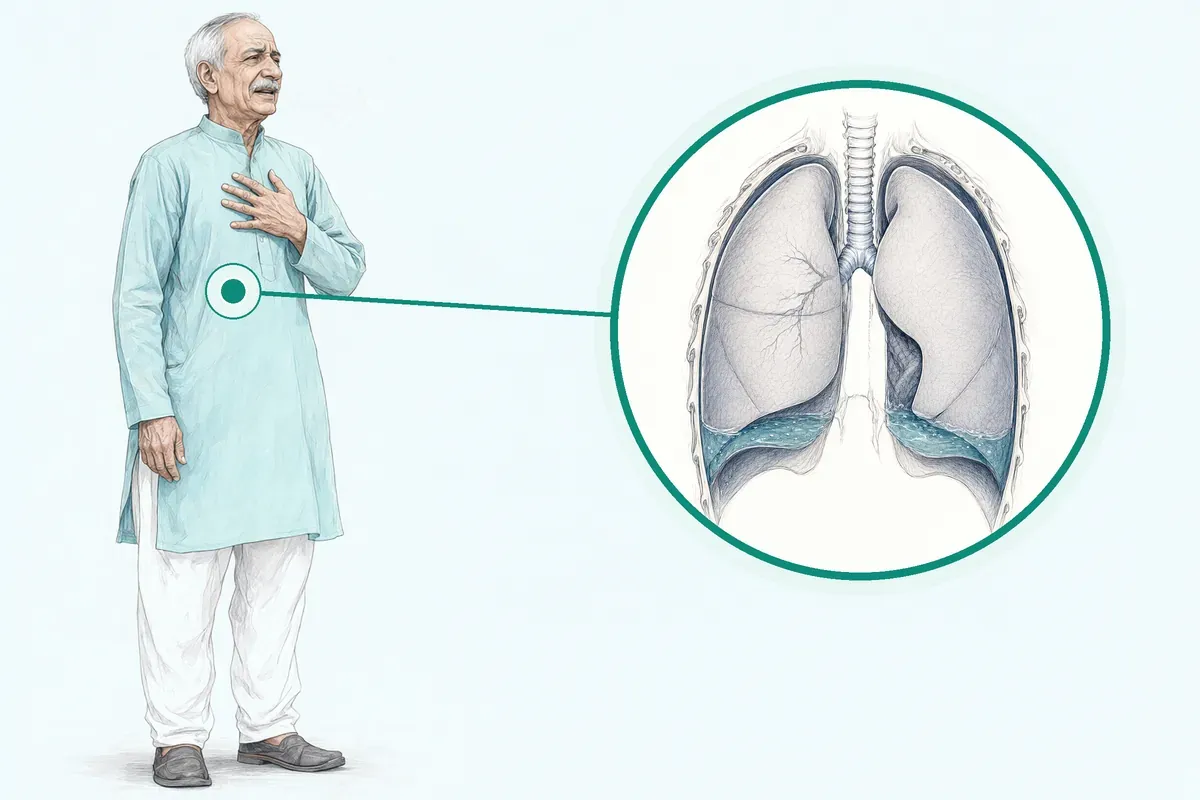
Extra fluid or air presses on your lung, making every breath an effort. You may also have a dry cough, fever, or pain when you breathe deeply. The cause could be infection, heart failure, injury, or cancer, so finding out why matters as much as draining it.
-
Shortness of breath
Climbing stairs or lying flat becomes hard because the lung cannot expand fully.
-
Chest discomfort
Sharp or dull pain on the affected side, worse with deep breaths or coughing.
-
Fever and fatigue
Common when the fluid is infected (empyema) or linked to pneumonia.
-
Unexplained weight loss
May signal an underlying malignancy that needs urgent investigation.
-
Recurrent episodes
Fluid comes back after drainage, needing a longer-term solution.
The most common causes of pleural effusion are congestive heart failure, cancer, pneumonia, and pulmonary embolism.
Why Jaipur Hospital
You worry that breathlessness will stop you from working or caring for your family, and you want to know the cost before you commit.
Your care plan
Your first steps with us
- 1 Call or visit OPD for a pulmonologist consultation
- 2 Get imaging and fluid tests done in-house
- 3 Undergo ultrasound-guided drainage or chest tube insertion
- 4 Recover with follow-up visits and clear home-care advice
How the operation is done
Procedures take 30 to 60 minutes under local anaesthesia with sedation. A needle or small tube is placed between the ribs using ultrasound guidance to drain fluid or air. A chest tube may stay for 2 to 5 days; wound heals in 1 to 2 weeks.
-
Therapeutic Thoracentesis
Ultrasound-guided needle drainage for immediate symptom relief; fluid sent for analysis.
-
Intercostal Chest Drain (ICD) Insertion
Tube placed for empyema, large pneumothorax or hemothorax; stays until lung re-expands.
-
Intrapleural Fibrinolytic Therapy
TPA or DNase instilled through the chest tube to break up loculated pus in empyema.
-
Medical Thoracoscopy (Pleuroscopy)
Scope inserted under local anaesthesia for visual biopsy and adhesiolysis in undiagnosed exudative effusion.
Why patients trust us
- 1991 Serving Jaipur for 35 years
- NABH Accredited hospital
- 1 Pulmonology consultants
- 24×7 Emergency, ICU and ambulance
- 25 Cashless schemes and insurers
What research shows about this treatment
-
Chest tube removal in 2 to 5 days
Studies report tube removal typically within this window after drainage starts.
-
Pleural effusion recurs in many patients
Even with drainage, effusions can be hard to control or come back due to malignancy.
-
Clinically symptomatic re-expansion pulmonary oedema in <1% of cases
Large-volume drainage rarely causes this serious complication when monitored.
If it is left untreated
Untreated fluid or air can lead to worsening breathlessness, lung collapse, or infection spreading to the bloodstream. Long-standing effusion may cause pleural thickening that traps the lung.
Life after treatment
Most people breathe easier within days and return to usual activities in a few weeks, with follow-up to watch for recurrence.
-
Easier breathing
Most people feel less breathless once the fluid or air is drained and the lung re-expands.
-
Return to routine
Usual activities often resume within 1 to 2 weeks after the tube is removed.
-
Ongoing monitoring
Follow-up scans and visits check that the lung stays expanded and the cause is managed.
-
Home drainage option
For recurrent malignant effusion, a tunneled catheter lets you drain at home with nurse support.

Frequently asked questions
Questions patients ask about pleural disease management
Which problems are assessed under pleural disease management?
In short
At Jaipur Hospital, our pulmonology team assesses pleural effusion, empyema, pneumothorax, haemothorax and malignant pleural effusion. These conditions involve excess fluid, pus or air in the pleural space around the lungs. The underlying causes range from infections and heart failure to cancer and trauma.
In detail
Pleural disease covers several distinct problems. Pleural effusion is a collection of fluid around the lungs, which may be watery (transudative) from heart failure or protein-rich (exudative) from pneumonia, cancer or pulmonary embolism.
Empyema is infected fluid or pus in the pleural space, often complicating pneumonia. Pneumothorax is air in the pleural cavity causing lung collapse. Haemothorax is blood in the pleural space, usually from trauma.
Malignant pleural effusion occurs when cancer cells are present in the fluid, most commonly from lung cancer in men and breast cancer in women. Mesothelioma, a primary pleural tumour, is associated with malignant effusion in more than 90 percent of cases. Our team uses clinical assessment, imaging and fluid analysis to identify the specific problem and its cause.
A common belief
Many people think pleural disease means only fluid on the lungs.
What actually happens
In fact, it includes infected fluid (empyema), air leaks (pneumothorax), blood (haemothorax) and cancer-related effusions. Each needs a different approach.
Your next step at Jaipur Hospital
If you have breathlessness, chest pain or a persistent cough, book a consultation with our pulmonology department. The doctor will review your history, examine you and order appropriate tests such as a chest X-ray or ultrasound. Most evaluations are completed within one to two visits.
How will the doctor decide which tests I need for pleural disease?
In short
The doctor at Jaipur Hospital decides tests based on your symptoms, examination findings and initial imaging. A chest X-ray is usually first. Ultrasound then confirms fluid location, volume and septations. Contrast-enhanced CT of the thorax is the gold-standard for pleural detail. If fluid is present, diagnostic thoracentesis with fluid analysis separates transudate from exudate and guides further investigation.
In detail
Ultrasound is significantly better than CT at identifying fibrous septations and is standard for guiding all pleural procedures. It avoids adhesions, targets pleural-based nodules, measures lung depth and uses colour Doppler to avoid intercostal vessels.
Pleural fluid cytology has 65 percent sensitivity for malignancy on first sampling. For undiagnosed exudative effusion, medical thoracoscopy provides visual biopsy with over 90 percent sensitivity. Blind pleural biopsies are no longer advocated due to lower yield. The choice depends on whether the effusion is transudative, exudative, loculated or suspicious for malignancy.
- Pleural fluid cytology has 65 percent sensitivity for malignancy on first sampling.
A common belief
Patients often ask for CT first, but ultrasound at the bedside is faster, avoids radiation and is superior for finding septations and safe needle sites.
What actually happens
Blind biopsies are outdated; image-guided or thoracoscopic biopsy gives over 90 percent diagnostic yield.
Your next step at Jaipur Hospital
Expect the doctor to order step-wise tests: X-ray, then ultrasound-guided thoracentesis for fluid analysis. If the cause remains unclear, CT thorax and possibly medical thoracoscopy are discussed. Most procedures take 30 to 60 minutes under local anaesthesia with sedation.
How soon should I expect improvement after pleural disease treatment?
In short
After pleural drainage or chest tube insertion at Jaipur Hospital, many patients feel less breathless within hours as the lung re-expands. Pain from the tube site improves over a few days. Full wound healing takes one to two weeks. If an underlying condition like infection or cancer is present, overall recovery depends on treating that cause.
In detail
Symptom relief from fluid or air removal is often immediate because the compressed lung can expand again. Evidence shows dyspnoea scores improve significantly after drainage.
Chest tube discomfort is common for the first few days; pain is reported in 5 to 39 percent of patients after thoracentesis and up to 15 percent after tube placement.
The tube usually stays in for 2 to 5 days until drainage decreases and the lung stays inflated. Re-expansion pulmonary oedema is rare, occurring in less than 1 percent of cases, but the team monitors for it.
For malignant effusions, median survival ranges from 3 to 12 months, so symptom control is the main goal. Your surgeon will explain what improvement timeline applies to your specific diagnosis.
A common belief
A common belief is that you feel completely normal right away.
What actually happens
In reality, if the effusion is due to cancer or a chronic infection, the underlying illness still limits your energy. Also, re-expansion pulmonary oedema, though rare, can cause new breathlessness if large volumes are removed too fast. That is why we monitor pleural pressure and stop drainage if you feel discomfort.
Your next step at Jaipur Hospital
You will be monitored in the ward with regular chest X-rays to confirm lung expansion. The tube is removed once drainage is low and no air leak persists.
Plan for a follow-up visit within one to two weeks after discharge to check the wound and review fluid or biopsy results. If breathlessness returns or you develop fever, contact the hospital sooner.
What is causing air or fluid around my lung?
In short
Air or fluid around the lung at Jaipur Hospital can have many causes. The most common causes of pleural effusion are congestive heart failure, cancer, pneumonia, and pulmonary embolism. Pneumothorax (air) often results from a ruptured bleb, trauma, or underlying lung disease. Your surgeon will identify the cause using imaging, fluid analysis, and sometimes biopsy.
In detail
Pleural effusion is classified as transudate (watery) or exudate (protein-rich). Transudates are usually due to heart failure, liver cirrhosis, or kidney disease. Exudates are caused by infections like pneumonia or tuberculosis, malignancies such as lung or breast cancer, pulmonary embolism, and autoimmune conditions.
Malignant pleural effusion is most often from lung cancer in men and breast cancer in women; mesothelioma is the primary pleural tumour in over 90 percent of cases. Pneumothorax can be primary spontaneous in young smokers, secondary from COPD or tuberculosis, or traumatic.
Ultrasound and CT help distinguish the cause. In some people, more than one cause exists simultaneously. The fluid itself may contain malignant cells or result from chemotherapy.
A common belief
Many patients think fluid always means cancer.
What actually happens
In fact, heart failure and pneumonia are very common causes. Also, a clear fluid (transudate) behaves differently from a protein-rich one (exudate), and the treatment changes completely. That is why we analyse the fluid before deciding on drainage or a chest tube.
Your next step at Jaipur Hospital
Your doctor will take a history, examine your chest, and order a chest X-ray or ultrasound. If fluid is present, a diagnostic thoracentesis is performed to send fluid for biochemistry, cytology, and microbiology. Further tests like CT thorax or medical thoracoscopy may be needed for a definite diagnosis.
How can I decide whether to seek advice about pleural tapping and ICD drainage?
In short
If you have breathlessness, chest heaviness or a cough that does not settle and a chest X-ray or ultrasound shows fluid around the lung, you should ask a pulmonologist at Jaipur Hospital about pleural tapping or intercostal chest drain insertion. These procedures relieve symptoms and give fluid for testing to find the cause.
In detail
Therapeutic thoracentesis uses an ultrasound-guided needle to remove fluid in thirty to sixty minutes under local anaesthesia with sedation. An intercostal chest drain is placed when fluid is infected, bloody or keeps coming back, or when a large pneumothorax needs continuous drainage.
Evidence shows ultrasound guidance reduces complications such as organ injury to below one percent. Fluid analysis separates transudate from exudate and guides further treatment. Your surgeon will choose the method based on fluid volume, thickness and whether the lung is trapped.
A common belief
Some people wait until they cannot lie flat.
What actually happens
Early drainage prevents lung collapse and makes diagnosis easier.
Your next step at Jaipur Hospital
You can attend the Pulmonology OPD for a clinical review and imaging. If a procedure is needed, it is usually done the same day or the next day. Hospital stay is two to six nights depending on drainage duration.
Which changes in breathing, chest symptoms, or oxygen levels need urgent help?
In short
Sudden worsening breathlessness, inability to speak in full sentences, oxygen saturation dropping below ninety percent on room air, sharp chest pain that increases with breathing, new confusion or drowsiness, and coughing up blood need immediate attention at Jaipur Hospital. These may signal tension pneumothorax, massive effusion, re-expansion pulmonary oedema or massive haemorrhage.
In detail
Re-expansion pulmonary oedema can occur within hours of rapid large-volume drainage and presents with hypoxia, cough and frothy sputum. Evidence shows clinically symptomatic re-expansion pulmonary oedema occurs in less than one percent of cases but is life-threatening.
Tension pneumothorax causes tracheal deviation, absent breath sounds and hemodynamic collapse. Massive haemorrhage from intercostal vessel injury is rare but can cause rapid blood loss. Persistent air leak beyond seven days with increasing subcutaneous emphysema also warrants urgent review. Any of these signs mean you should call the emergency number or attend the 24×7 emergency department without delay.
A common belief
Some people wait to see if it passes.
What actually happens
Re-expansion pulmonary oedema and tension pneumothorax worsen in minutes and can be fatal without immediate treatment.
Your next step at Jaipur Hospital
If you are an inpatient, press the call bell. If you are at home after a procedure, call the hospital emergency line or come straight to the emergency department. Keep your discharge summary and medication list handy.
Which symptoms should prompt me to see a specialist for pleural disease?
In short
At Jaipur Hospital, you should see a specialist if you have breathlessness that worsens with activity or lying flat, chest pain that is sharp and worse on breathing, a persistent dry cough, fever with chest symptoms, or unexplained weight loss. Some people have no symptoms and the effusion is found on a chest X-ray done for another reason.
In detail
Pleural effusion symptoms include shortness of breath, chest discomfort, and cough. The breathlessness occurs because excess fluid limits lung expansion. Chest pain is typically pleuritic, meaning it worsens with deep breaths or coughing. Fever suggests an infectious cause such as empyema or parapneumonic effusion.
Weight loss and fatigue may point to a malignant cause. In malignant pleural effusion, disabling dyspnoea is common and often occurs with a short life expectancy. Some patients have unrelated symptoms from the underlying disease, such as heart failure or cancer.
A physical examination by a doctor includes listening to the lungs for reduced breath sounds or a stony dull note on percussion. These findings guide the need for imaging and further tests.
A common belief
People often wait until they cannot breathe comfortably.
What actually happens
But early signs like mild breathlessness on exertion or a cough that does not go away can be the first clues. Fever with chest pain suggests infection needing urgent drainage. Weight loss may signal cancer.
Your next step at Jaipur Hospital
Do not ignore persistent breathlessness or chest pain. Book an appointment with our pulmonology department for a clinical evaluation. The doctor will examine you and may order a chest X-ray or ultrasound on the same day. Early assessment helps identify the cause and start treatment promptly.
How will the results change my care plan for pleural disease?
In short
At Jaipur Hospital, test results directly guide your treatment. Transudative fluid points to heart failure or liver disease, managed by treating the underlying condition. Exudative fluid suggests infection, cancer or autoimmune disease, needing antibiotics, drainage or oncology referral. Malignant cells confirm malignant pleural effusion, leading to pleurodesis or indwelling catheter discussion. Empyema requires chest tube drainage and possibly fibrinolytic therapy.
In detail
The fluid analysis is the pivot point. Light's criteria (fluid protein, LDH, glucose, pH) classify the effusion. Transudates are treated by optimising heart failure, renal or hepatic therapy.
Exudates with low pH, low glucose and high LDH suggest complicated parapneumonic effusion or empyema, requiring urgent intercostal chest drain insertion and possibly intrapleural fibrinolytic therapy (TPA/DNase) for loculated collections. Positive cytology or biopsy confirms malignant pleural effusion.
Options include therapeutic thoracentesis for symptom relief, talc pleurodesis via chest tube or thoracoscopy, or indwelling pleural catheter for ambulatory drainage. In studies, indwelling catheters reduced hospital days compared to talc pleurodesis.
For undiagnosed exudative effusion, medical thoracoscopy provides visual biopsy with over 90 percent sensitivity. Pneumothorax size and symptoms decide between observation, aspiration or chest tube insertion. Your surgeon will explain the recommended path based on your specific results.
A common belief
Patients often expect the same treatment for any fluid.
What actually happens
But a transudate from heart failure improves with diuretics, not drainage. An empyema needs a tube and possibly clot-busting drugs. A malignant effusion needs a strategy to prevent recurrence. The fluid chemistry and cytology tell us which path to take.
Your next step at Jaipur Hospital
Once results are available, usually within 24 to 48 hours for fluid analysis and imaging, the doctor will discuss the treatment plan with you and your attendant. Procedures like chest tube insertion or thoracoscopy are scheduled based on urgency. Hospital stay ranges from 2 to 6 nights depending on drainage duration.
What should I do if symptoms persist or worsen after pleural treatment?
In short
If breathlessness, chest pain, fever, or cough return or get worse after pleural treatment at Jaipur Hospital, contact the hospital immediately or visit the 24-hour emergency department. These symptoms may indicate a recurrent effusion, persistent air leak, infection, or re-expansion pulmonary oedema. Early review prevents complications.
In detail
Persistent air leak is defined as bubbles in the water seal lasting more than three to seven days. Re-expansion pulmonary oedema typically appears within minutes to hours after large-volume drainage, causing sudden dyspnoea, cough, and hypoxia.
Infection at the drain site is rare under sterile conditions but can occur. Evidence shows clinically symptomatic re-expansion pulmonary oedema occurs in less than 1 percent of cases, while post-procedure pain is reported in 5 to 39 percent of patients.
If you had a chest tube removed and then develop increasing breathlessness or a new fever, the lung may not have stayed expanded or an empyema could be developing. Your attendant should bring you to the emergency department for a chest X-ray and clinical assessment without delay.
A common belief
Some patients think mild breathlessness is normal for weeks.
What actually happens
It is not. A persistent air leak beyond a week, new fever, or sudden shortness of breath after drainage needs urgent imaging. Waiting can turn a small problem into a longer hospital stay.
Your next step at Jaipur Hospital
Keep the discharge summary and any prescribed medicines handy. Do not wait for the next scheduled follow-up if you feel worse. The emergency team at Jaipur Hospital is available 24 hours a day with on-site imaging and laboratory support. Call the hospital numbers or come directly to the emergency entrance.
Will the fluid need testing or a biopsy?
In short
Yes, fluid removed during thoracentesis at Jaipur Hospital is routinely sent for testing to find the cause. Tests include protein and LDH levels to separate transudate from exudate, cell counts, microbiology for infection, and cytology for cancer cells. If cytology is negative but suspicion remains, a pleural biopsy via medical thoracoscopy or image-guided needle may be recommended.
In detail
Pleural fluid analysis is the first step. It differentiates transudate from exudate, which narrows the diagnosis. Cytology detects malignant cells with about 65 percent sensitivity on the first sample; for mesothelioma it is lower, around 20 to 33 percent.
Blind pleural biopsies are no longer recommended due to low yield. Medical thoracoscopy provides direct vision and biopsy with over 90 percent sensitivity for malignant pleural effusion. Ultrasound-guided pleural biopsy is a valid alternative with 94 percent yield in frail patients.
Biomarkers like mesothelin in serum or fluid can support a malignancy diagnosis. Your surgeon decides on biopsy based on fluid results, imaging findings, and your fitness for the procedure.
- Ultrasound-guided pleural biopsy is a valid alternative with 94 percent yield in frail patients.
A common belief
A common misconception is that one fluid test rules out cancer.
What actually happens
Cytology misses about a third of malignancies on the first try, and even more for mesothelioma. That is why guidelines prefer a tissue biopsy when suspicion is high. We do not do blind biopsies anymore; we use ultrasound or thoracoscopy to target the abnormal area directly.
Your next step at Jaipur Hospital
After a diagnostic tap, you will get preliminary fluid results within a few hours; cytology and culture take 2 to 5 days. If a biopsy is needed, the team will schedule medical thoracoscopy or an image-guided biopsy, usually within a week. Both are done under local anaesthesia with sedation. Plan for a short hospital stay.
FAQ library
Questions and answers about pleural disease management
When you contact us
Get your assessment and plan
Contact Jaipur Hospital for a pulmonologist consultation, in-house tests, a plain-word explanation of options, a cost estimate and help with cashless schemes.
- A consultation with a pulmonologist (chest physician) in the OPD
- Your scans and reports reviewed, with tests done in-house if you need them (laboratory, X-ray and imaging)
- Your options explained in plain words, including whether you need surgery at all
- A cost estimate before you decide
- Help with cashless treatment under RGHS, MAA Yojana, CGHS and empanelled insurers and TPAs
- Follow-up visits after your treatment
Overview
Pleural Disease Management: what to know
What is pleural disease
- Pleura
- Two thin membranes lining lungs and chest cavity
- Normal fluid
- Small amount works as natural lubricant
- Pleural effusion
- Buildup of excess fluid around the lungs
- Empyema
- Pus collects in the pleural space
- Pneumothorax
- Air leaks into the space and can collapse the lung
The pleura are two thin membranes that line your lungs and the inside of your chest cavity. Normally, a small amount of fluid sits between these layers and works as a natural lubricant so your lungs move smoothly when you breathe. Pleural disease means something has upset this balance.
The most common problem is pleural effusion, a buildup of excess fluid around the lungs. Other conditions we treat include empyema, where pus collects in the pleural space, and pneumothorax, where air leaks into the space and can collapse the lung. These conditions can develop quickly or slowly depending on the cause.
Symptoms of pleural effusion and when to seek urgent care
- Sudden severe breathlessness
- Sharp chest pain with breathing
- Feeling faint
- Coughing up blood
- High fever with chills
Many people with a small pleural effusion have no symptoms at all and find out only when they have a chest X-ray for another reason.
When symptoms appear, they often include:
- Shortness of breath
- Chest pain that gets worse with breathing or coughing
- A dry cough
- Sometimes fever
In malignant pleural effusion, disabling breathlessness is common.
Seek care the same day if you suddenly feel very short of breath, have sharp chest pain with breathing, feel faint, cough up blood, or develop a high fever with chills. These can signal a large effusion, a pneumothorax, or an infection such as empyema that needs immediate drainage.
Causes of pleural disease and who is at higher risk
Pleural effusion has many causes, and some people have more than one. Doctors group the fluid into two types: transudative (watery) and exudative (protein-rich). The most common causes of transudative effusion are congestive heart failure, liver disease (cirrhosis), and kidney disease (nephrotic syndrome). Common causes of exudative effusion include pneumonia, cancer, pulmonary embolism, and tuberculosis.
Less common causes include:
- Autoimmune diseases
- Pancreatitis
- Reactions to certain medicines
Malignant pleural effusion is most often caused by lung cancer in men and breast cancer in women. Mesothelioma, a primary tumour of the pleura, is linked to asbestos exposure and causes malignant effusion in more than 90% of cases. Primary spontaneous pneumothorax usually affects young, otherwise healthy people, and smoking is the main risk factor.
Empyema often follows pneumonia that was not fully treated. Having any condition that causes fluid overload or inflammation in the chest raises your risk.
How pleural disease is diagnosed: examination and tests
- 65% Pleural fluid cytology first sample
- above 90% Medical thoracoscopy sensitivity
- 94% Ultrasound-guided biopsy yield
Your doctor will ask about your medical history, other illnesses, and when your symptoms started. A physical examination includes listening to your lungs with a stethoscope.
Imaging is the next step. A chest X-ray is usually the first test. Ultrasound of the chest is significantly better than CT at identifying fibrous septations and helps guide needle placement. Contrast-enhanced CT of the thorax is the gold-standard imaging modality for the pleura and shows thickening, nodules, and the extent of disease.
To find the cause, fluid is removed with a needle (thoracentesis or pleural tap) and sent to the laboratory.
The fluid is tested for:
- Protein
- Lactate dehydrogenase (LDH)
- Cell counts
- Microbiology
- Cytology
Pleural fluid cytology picks up malignant cells on the first sample in about 65% of cases, but the yield is lower for mesothelioma. If cytology is negative and the cause remains unclear, image-guided pleural biopsy or medical thoracoscopy (pleuroscopy) is advised.
Medical thoracoscopy allows direct visual inspection and biopsy under local anaesthesia with sedation, with a diagnostic sensitivity above 90% for malignant pleural effusion. Ultrasound-guided pleural biopsy is a valid alternative for frail patients or those with tethered lungs, with a reported yield of 94%.
Non-surgical treatment options for pleural disease and when surgery is advised
- Therapeutic thoracentesis Quick fluid relief for symptomatic effusion
- Chest drain insertion For empyema, hemothorax, or large pneumothorax
- Fibrinolytic therapy Breaks up trapped fluid pockets
- Indwelling pleural catheter Home drainage for recurrent malignant effusion
- Medical thoracoscopy with talc Diagnosis and pleurodesis in one procedure
- Surgery (VATS) When other treatments fail or lung is trapped
Treatment depends on the underlying cause and how much the fluid or air is affecting your breathing.
For a symptomatic pleural effusion, therapeutic thoracentesis (ultrasound-guided fluid removal) gives quick relief. If the effusion is large, drainage is done slowly while monitoring pleural pressure to avoid re-expansion pulmonary oedema, a rare but serious complication.
For empyema, hemothorax, or a large pneumothorax, an intercostal chest drain (tube) is inserted under local anaesthesia with sedation. The tube stays in place for 2 to 5 days until the lung re-expands and drainage stops.
If the fluid is loculated (trapped in pockets), intrapleural fibrinolytic therapy (TPA/DNase) can be given through the chest tube to break up the septations and improve drainage.
For recurrent malignant pleural effusion, an indwelling tunneled pleural catheter (IPC) can be placed. This silicone tube is tunnelled under the skin and allows you or an attendant to drain fluid at home, reducing hospital stay. In studies, IPCs gave similar breathlessness relief to talc pleurodesis at 6 weeks, with significantly fewer hospital days.
Medical thoracoscopy (pleuroscopy) is advised when the diagnosis is still unclear after fluid analysis, or when adhesions need to be divided (adhesiolysis) to allow the lung to expand. During thoracoscopy, talc poudrage (spraying sterile talc) can be done at the same time to stick the pleural layers together and prevent fluid from coming back.
Talc is the most commonly used agent for pleurodesis, and graded (calibrated) talc is used to minimise the risk of acute respiratory distress syndrome.
Surgery (video-assisted thoracic surgery or VATS) is considered when drainage and pleurodesis have failed, when the lung cannot expand (trapped lung), or for definitive management of recurrent pneumothorax. Your surgeon will discuss which approach suits your condition and overall health.
Preparing for pleural disease management surgery
Do
- Bring recent imaging reports
- Bring your medicine list
- Bring an attendant
- Wear loose clothing
- Fast for at least six hours
Before any pleural procedure, your surgical team will review your medical history and current medicines. Blood tests are usually done to check clotting and kidney function. If you take blood thinners such as warfarin, apixaban or clopidogrel, your doctor will discuss when to stop them; aspirin is generally considered safe to continue.
For urgent procedures where anticoagulation cannot be stopped, the risk and benefit are assessed together with you. You will need to fast for at least six hours before the procedure because sedation is used. Wear loose clothing and bring any recent imaging reports, your medicine list and an attendant who can stay with you during the hospital stay.
The team will confirm the side of the chest to be treated and mark the site after an ultrasound scan.
How pleural disease management surgery is done
- Therapeutic thoracentesis Ultrasound-guided needle drainage under local anaesthesia with sedation, about 30 minutes
- Intrapleural fibrinolytic therapy TPA and DNase instilled through chest tube to break up loculated pus in empyema
- Indwelling tunneled pleural catheter Silicone tube tunnelled under skin for home drainage of recurrent malignant effusion
The procedure you have depends on the type and cause of the pleural problem. Your surgeon will advise which technique suits you. Therapeutic thoracentesis is an ultrasound-guided needle drainage done under local anaesthesia with sedation; it takes about 30 minutes and is used for symptomatic fluid relief.
Intercostal chest drain (ICD) insertion places a tube through the chest wall under local anaesthesia with sedation to drain empyema, hemothorax or a large pneumothorax; the tube stays in for two to five days. Intrapleural fibrinolytic therapy instills tissue plasminogen activator and DNase through an existing chest tube to break up loculated pus in empyema.
Medical thoracoscopy (pleuroscopy) uses a rigid or semi-rigid scope inserted through a small incision under local anaesthesia with sedation or general anaesthesia; it allows direct vision, biopsy and adhesiolysis for undiagnosed exudative effusion. On the day, you lie on your side with the affected side up.
The skin is cleaned, local anaesthetic is injected, and the chosen instrument is introduced under ultrasound guidance to avoid vessels and adhesions. The procedure typically lasts 30 to 60 minutes. If talc pleurodesis is needed, sterile graded talc is insufflated under vision at the end of thoracoscopy. An indwelling tunneled pleural catheter may be placed during the same sitting for recurrent malignant effusion.
Risks and possible complications of pleural disease management
- 5 to 39 percent Pain during or after thoracentesis
- 8 percent Pain during chest tube placement
- 15.6 percent Delayed pain after chest tube
- about 2 percent Re-expansion pulmonary oedema after thoracoscopy
- around 1.5 percent Overall major complication rate
Pleural procedures are generally safe. In studies, pain during or after thoracentesis is reported in 5 to 39 percent of patients. For chest tube placement, 8 percent report pain during the procedure and 15.6 percent report delayed pain. Significant bleeding is uncommon; most post-procedural bleeding is superficial and controlled with external pressure. Massive haemorrhage can occur but is rare.
Injury to the liver, spleen or diaphragm has been reported in 0.3 percent of thoracenteses and 0.6 percent of chest tube placements. Infection at the procedure site is rare when sterile technique is used; with proper care of indwelling catheters the infection rate remains low.
Re-expansion pulmonary oedema is reported in about 2 percent of patients after medical thoracoscopy; clinically symptomatic cases occur in less than 1 percent of large-volume drainage. Persistent air leak lasting three to seven days can occur after pleural procedures. Malignant seeding of the entry tract is a particular concern in mesothelioma.
Temporary rise in arterial carbon dioxide and reduced lung function may happen in the 48 hours after thoracoscopy, so patients with baseline hypercapnia are monitored closely. Overall major complication rates in studies are around 1.5 percent.
Recovery and aftercare after pleural disease management
After thoracentesis you are usually advised to take it easy for two days. If a chest tube is inserted, you stay in hospital for two to six nights until drainage decreases and the lung remains expanded; the tube is typically removed in two to five days. Wound healing takes one to two weeks.
After medical thoracoscopy or VATS, a few days in hospital are common; open thoracotomy requires about a week. Indwelling pleural catheter placement often allows same-day discharge. Before discharge you receive instructions on wound care, breathing exercises and when to resume normal activity.
Your attendant should watch for:
- Fever
- Increasing chest pain
- Sudden breathlessness
- Redness or discharge at the wound site
- Persistent air bubbling in the drainage bottle if a tube is still in place
Contact the hospital or visit the 24×7 emergency department if any of these occur. Follow-up chest X-rays are usually done after treatment.
Results of pleural disease management: how well it works and recurrence
In studies of malignant pleural effusion, both indwelling pleural catheters and talc pleurodesis improve breathlessness over the first six weeks with no significant difference. At six months, catheter patients reported greater improvement in dyspnoea scores. Pleurodesis success rates in large randomised trials remain under 80 percent and are likely lower in routine practice.
Talc poudrage during thoracoscopy and talc slurry through a chest tube have shown similar success overall, though a subgroup analysis suggested better results with poudrage for lung and breast cancer. A recent meta-analysis found talc poudrage superior to slurry.
Indwelling catheters reduce total hospital days from procedure to death or 12 months (median 10 versus 12 days) and cut effusion-related admissions (median 1 versus 4 days). For primary spontaneous pneumothorax, definitive treatment with pleurodesis after recurrence lowers further recurrence risk. Pleural effusion can recur, especially when caused by malignancy. Median survival with malignant pleural effusion ranges from three to 12 months. Your outlook depends on the underlying cause.
Cost and insurance for pleural disease management in Jaipur
The typical price range for pleural procedures at Jaipur Hospital is ₹30,000 to ₹1,20,000. The final cost depends on the type of procedure (thoracentesis, chest tube, fibrinolytic therapy or thoracoscopy), length of hospital stay, whether pleurodesis or an indwelling catheter is needed, and any additional investigations or ICU care.
Jaipur Hospital is a NABH-accredited multispeciality hospital established in 1991 with modular operation theatres, intensive care, in-house laboratory and imaging, and 24×7 emergency and ambulance services.
The hospital accepts:
- RGHS
- MAA Yojana
- CGHS
- A panel of private insurers and TPAs
Ask the admissions desk for an estimate based on your planned procedure and insurance coverage.