

Pulmonology · Jaipur Hospital
Pulmonary Vascular Disease Care in Jaipur
When breathlessness or chest discomfort limits your day, our team offers evaluation, scans, and an organised medical plan.
- Serving Jaipur since 1991 (35 years)
- NABH accredited
- 24×7 emergency & ICU
- Cashless: RGHS, MAA Yojana, CGHS + 22 insurers
- Treatment length
- Acute PE: 5 to 10 days anticoagulationPH: Lifelong medical therapy
- Anaesthesia
- Not needed (medical)General anaesthesia (PTE surgery for CTEPH)
- Recovery
- Acute PE: 3 to 6 months anticoagulationPH: Chronic management · PTE surgery: 8 to 12 weeks
- Price
- ₹50,000 to ₹3,00,000 (acute)₹20,00,000+ (PTE surgery at referral centre)typical range in India; ask us for an estimate
Struggling to catch your breath
You feel breathless and tired, but answers remain unclear
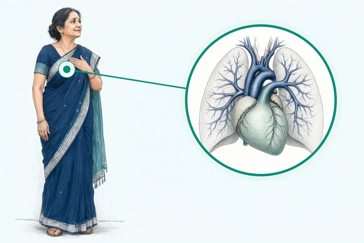
Sudden or lingering shortness of breath can make daily steps difficult. You may wonder if your symptoms come from your heart, your lungs, or a blood clot, leaving you unsure of what steps to take next.
-
Unexplained breathlessness
Shortness of breath during simple tasks can happen even when resting tests look unremarkable.
-
Lingering chest pain
Discomfort or heaviness in the chest is sometimes misread as muscle strain rather than vascular strain.
-
Fatigue after a clot
Breathlessness can continue for months after a deep vein clot or acute pulmonary event.
Disregarding patient complaints of unexplained dyspnea as anxiety or hyperventilation is a common pitfall in management.
Why Jaipur Hospital
You may worry about unexpected hospital costs, missing work, or finding a clear diagnosis when breathing feels difficult.
Your care plan
Your first steps with us
- 1 Visit our OPD for a pulmonology evaluation.
- 2 Complete in-house blood tests, imaging, or heart assessments.
- 3 Discuss your medical options with your physician.
- 4 Follow your prescribed medical plan and schedule reviews.
How pulmonary vascular conditions are evaluated
Your physician will review your clinical history, assess clot risks, and conduct targeted imaging or hemodynamic evaluations to determine an appropriate medical or procedural approach.
-
CT Pulmonary Angiography (CTPA)
This imaging option provides detailed views of the pulmonary arteries to assess acute clots, as your physician will discuss.
-
Right Heart Catheterisation (RHC)
This procedure measures pressures inside the heart and pulmonary arteries directly, an option your physician will discuss.
-
Anticoagulation / Thrombolysis
Blood-thinning medications or clot-dissolving therapies serve as medical options your physician will discuss based on clinical stability.
-
Pulmonary Vasodilator Therapy
Targeted medications help relax constricted pulmonary vessels in pulmonary arterial hypertension, which your physician will discuss.
Why patients trust us
- 1991 Serving Jaipur for 35 years
- NABH Accredited hospital
- 1 Pulmonology consultants
- 24×7 Emergency, ICU and ambulance
- 25 Cashless schemes and insurers
Clinical findings in vascular care
-
1.1%
In a study of 4832 patients with acute pulmonary embolism, major bleeding occurred in 1.1% of patients receiving rivaroxaban.
-
3.3%
In a trial subgroup with right ventricular dysfunction, the recurrent venous thromboembolism rate was 3.3% with edoxaban.
-
20% to 24%
Applying structured clinical algorithms reduced the need for follow-up echocardiography investigation to 20% to 24% of tested patients.
If it is left untreated
Leaving pulmonary vascular conditions unaddressed can lead to progressive strain on the right heart, persistent exercise limitation, or recurrent blood clots. Failing to initiate timely diagnostic workup or appropriate anticoagulation in suspected pulmonary embolism can result in significant clinical complications.
Life with managed care
With structured medical therapy and routine reviews, most patients find clearer daily steps and a defined management routine.
-
Structured medication routine
Most people establish an organised schedule for oral anticoagulation or vasodilator therapy from their local chemist.
-
Clear activity pacing
Patients typically plan daily work and walking according to medical advice and follow-up guidance.
-
Regular OPD monitoring
Routine visits allow your attendant and physician to review scans, reports, and adjust medical plans.
Frequently asked questions
Questions patients ask about pulmonary vascular disease care
Which problems are assessed under pulmonary vascular disease?
In short
Pulmonary vascular disease care at Jaipur Hospital covers acute pulmonary embolism, pulmonary hypertension and chronic thromboembolic pulmonary hypertension. These conditions affect the blood vessels of the lungs and can cause breathlessness, chest discomfort and reduced exercise capacity.
In detail
Acute pulmonary embolism is a sudden blockage in a lung artery, usually from a blood clot that travels from the legs. Pulmonary hypertension means the pressure in the lung arteries is higher than normal, which makes the right side of the heart work harder.
Chronic thromboembolic pulmonary hypertension develops when old clots do not dissolve and leave scar tissue that narrows the arteries. Right heart catheterisation gives the definitive haemodynamic diagnosis for pulmonary hypertension, while CT pulmonary angiography is the key imaging test for acute pulmonary embolism. The hospital follows international classification that groups these diseases by shared pathology and treatment approach.
A common belief
People often think breathlessness is only a lung or heart problem.
What actually happens
In reality, the lung blood vessels themselves can be the primary site of disease, needing specific tests like right heart catheterisation to confirm.
Your next step at Jaipur Hospital
If you have been diagnosed with any of these conditions or have persistent unexplained breathlessness, you can book a consultation with our pulmonology team. Bring any previous imaging, blood test results and a list of current medicines.
The doctor will review your history, decide which tests are needed and discuss a management plan. To schedule an appointment, contact Jaipur Hospital.
How will the doctor decide which tests I need?
In short
The doctor at Jaipur Hospital decides tests based on your symptoms, clinical examination findings, risk factors and the suspected diagnosis. The approach follows a step-by-step pathway: clinical assessment first, then non-invasive tests, then invasive confirmation if needed.
In detail
If acute pulmonary embolism is suspected, the doctor uses clinical probability scores such as Wells or simplified PESI, then D-dimer and CT pulmonary angiography. For pulmonary hypertension, echocardiography is the initial screening test to estimate pulmonary artery pressure and assess right heart function.
If echocardiography shows intermediate or high probability of pulmonary hypertension, right heart catheterisation is recommended for definitive haemodynamic diagnosis. For chronic thromboembolic pulmonary hypertension, guidelines recommend perfusion imaging such as VQ SPECT, dual-energy CT or MR perfusion mapping combined with echocardiography.
The InShape algorithms use CTPA reading of the index pulmonary embolism, ECG and NT-proBNP to decide who needs echocardiographic screening. The doctor also considers your bleeding risk, renal function and comorbidities before ordering contrast studies or invasive tests.
A common belief
Patients sometimes ask for every test at once.
What actually happens
Guidelines show a structured pathway avoids unnecessary tests: echocardiography screens for pulmonary hypertension, CT pulmonary angiography diagnoses acute pulmonary embolism, and right heart catheterisation confirms pulmonary hypertension haemodynamically. Each test has a specific role.
Your next step at Jaipur Hospital
After the consultation, the team will schedule the required tests, usually within a few days. You may need blood tests before contrast imaging. Right heart catheterisation requires a short hospital stay. The doctor will explain the purpose of each test and any preparation needed. To start the diagnostic pathway, book a consultation at Jaipur Hospital.
How soon should I expect improvement after the chosen treatment for pulmonary vascular disease?
In short
Improvement timelines depend on the condition and treatment. For acute pulmonary embolism on anticoagulation, breathlessness often eases within days to weeks, while full exercise recovery may take three to six months. For pulmonary arterial hypertension on targeted therapy, functional class improvement typically appears over three to six months. After pulmonary endarterectomy for chronic thromboembolic pulmonary hypertension, haemodynamic improvement is immediate but functional recovery takes eight to twelve weeks.
In detail
In acute pulmonary embolism, anticoagulation prevents new clots while the body gradually dissolves the existing clot; symptom relief correlates with clot resolution and right ventricular recovery. Studies show most patients on direct oral anticoagulants achieve similar recurrence and bleeding outcomes to standard therapy.
For pulmonary arterial hypertension, endothelin receptor antagonists, phosphodiesterase-5 inhibitors and prostacyclin analogues improve exercise capacity and haemodynamics over months; combination therapy may accelerate response.
After pulmonary endarterectomy, patients experience significant improvement in WHO functional class, exercise capacity and quality of life, with median follow-up showing sustained benefit. Right heart catheterisation at follow-up confirms haemodynamic normalisation. Rehabilitation programmes during recovery enhance functional gains.
Persistent symptoms beyond expected timelines prompt reassessment for residual disease, recurrent embolism or alternative diagnoses such as left heart disease.
A common belief
Many patients expect instant relief.
What actually happens
In pulmonary arterial hypertension, medicines work slowly to remodel vessels. After surgery, the heart and lungs need time to adapt. Persistent breathlessness at three months does not mean treatment failed; it signals the need for re-evaluation.
Your next step at Jaipur Hospital
Your surgeon will set specific milestones based on your baseline severity and treatment type. Follow-up echocardiography at three months and six months tracks progress. If symptoms do not improve as expected, further imaging or right heart catheterisation may be needed.
How can I decide whether to seek advice about pulmonary embolism management?
In short
Seek advice promptly if you have sudden breathlessness, chest pain that worsens with breathing, fainting, coughing blood, or leg swelling and pain. Jaipur Hospital has a 24-hour emergency department for immediate assessment. Even if symptoms are mild or intermittent, a history of recent surgery, immobility, cancer, pregnancy or prior clot raises the likelihood of pulmonary embolism.
In detail
Clinical suspicion is the first step. Guidelines list Wells score, revised Geneva score and PERC rule to estimate probability. If probability is not low, D-dimer testing or immediate CT pulmonary angiography is indicated. Low-risk patients with negative PERC criteria may not need testing.
However, disregarding unexplained dyspnoea as anxiety or attributing chest pain to musculoskeletal causes are common pitfalls that delay diagnosis. Starting therapeutic anticoagulation before confirmatory imaging is recommended when clinical probability is intermediate or high.
If you have had a previous pulmonary embolism and develop new or worsening symptoms, re-evaluation is necessary to exclude recurrence or chronic thromboembolic disease. The 2019 ESC guidelines recommend follow-up echocardiogram at 3 to 6 months for persistent dyspnoea or CTEPH risk factors.
A common belief
Many people dismiss breathlessness as anxiety or being out of shape.
What actually happens
Chest pain is often blamed on muscle strain. These delays can be fatal. Also, a normal D-dimer does not rule out PE in high-probability cases; imaging is needed. And if you have had PE before, new symptoms are not just a recurrence; they could signal chronic thromboembolic disease needing a different workup.
Your next step at Jaipur Hospital
Do not self-diagnose or wait for symptoms to resolve. Call the emergency number or visit the OPD at Jaipur Hospital. Early evaluation prevents complications.
What are Pulmonary Vascular Diseases?
In short
Pulmonary vascular diseases are conditions that affect the blood vessels of the lungs, including pulmonary embolism, pulmonary hypertension, and chronic thromboembolic pulmonary hypertension. Jaipur Hospital provides diagnosis and management for these conditions using imaging, haemodynamic assessment, and targeted medical or surgical therapy.
In detail
Pulmonary embolism is acute blockage of pulmonary arteries by thrombus, diagnosed by CT pulmonary angiography. Pulmonary hypertension is a mean pulmonary artery pressure of 25 mmHg or more at rest on right heart catheterisation, classified into five groups sharing pathological and haemodynamic features.
Group 1 is pulmonary arterial hypertension, defined by pre-capillary pulmonary hypertension with pulmonary vascular resistance of three Wood units or more. Group 4 is chronic thromboembolic pulmonary hypertension, caused by organised thromboembolic obstruction after acute pulmonary embolism.
Acute pulmonary embolism management includes anticoagulation with low molecular weight heparin or direct oral anticoagulants, thrombolysis for massive pulmonary embolism with shock, and inferior vena cava filters in selected cases.
Pulmonary arterial hypertension therapy includes endothelin receptor antagonists, phosphodiesterase-5 inhibitors, prostacyclin analogues, and soluble guanylate cyclase stimulators, often in combination. Chronic thromboembolic pulmonary hypertension may be treated with pulmonary endarterectomy, balloon pulmonary angioplasty, or medical therapy. Right heart catheterisation remains essential for definitive diagnosis of pulmonary hypertension.
A common belief
People often confuse pulmonary hypertension with high blood pressure in the body.
What actually happens
Pulmonary hypertension is high pressure in the lung arteries specifically, measured by right heart catheterisation. It has five distinct groups with different causes and treatments. Acute pulmonary embolism is a medical emergency, while chronic thromboembolic pulmonary hypertension develops months later from organised clots. Each requires a different diagnostic pathway and treatment strategy.
Your next step at Jaipur Hospital
If you have symptoms suggesting a pulmonary vascular disease, book a consultation at Jaipur Hospital. Your surgeon will arrange the appropriate imaging and haemodynamic tests, explain the diagnosis, and discuss a management plan tailored to your condition.
Which changes in breathing, chest symptoms, or oxygen levels need urgent help?
In short
Seek urgent help for sudden severe breathlessness, chest pain that worsens with breathing, coughing up blood, fainting or dizziness, cold sweats, or oxygen saturation below 90 percent at rest. Jaipur Hospital operates a 24-hour emergency department equipped for rapid assessment of acute pulmonary embolism and pulmonary hypertension crises.
In detail
Acute massive pulmonary embolism presents with haemodynamic instability, shock, or cardiac arrest. Submassive pulmonary embolism shows right ventricular dysfunction on imaging or elevated troponin without haemodynamic collapse. Both require immediate hospital admission.
In pulmonary hypertension, acute right heart failure manifests as rapid worsening of breathlessness, syncope, peripheral oedema, and rising jugular venous pressure. Oxygen desaturation at rest or with minimal exertion signals significant ventilation-perfusion mismatch or right-to-left shunting.
Guidelines emphasise that failing to initiate therapeutic anticoagulation in suspected pulmonary embolism before confirmatory imaging is a common pitfall. Similarly, dismissing unexplained dyspnoea as anxiety or attributing chest pain to musculoskeletal causes delays life-saving treatment.
Pulse oximetry is a simple screening tool but normal saturation does not exclude pulmonary embolism. Clinical probability scores such as Wells or revised Geneva guide the need for D-dimer or immediate CT pulmonary angiography.
A common belief
Many people wait, thinking breathlessness is anxiety, a panic attack, or a chest infection.
What actually happens
Guidelines show that dismissing unexplained dyspnoea as anxiety is a common pitfall that delays pulmonary embolism diagnosis. Chest pain from pulmonary embolism is often pleuritic and mistaken for muscle strain. Normal oxygen saturation does not rule out pulmonary embolism. Clinical probability scores, not symptoms alone, should trigger immediate imaging and anticoagulation.
Your next step at Jaipur Hospital
If you experience any of these symptoms, call an ambulance or go to the nearest emergency department. Jaipur Hospital provides 24-hour emergency care with in-house CT pulmonary angiography, echocardiography, and intensive care. Your surgeon will be notified for immediate assessment if pulmonary vascular disease is suspected.
Which symptoms should prompt me to see a specialist?
In short
You should see a specialist at Jaipur Hospital if you have new or worsening breathlessness on exertion, unexplained chest discomfort, fainting or near-fainting episodes, swelling of the ankles, or a persistent cough. These symptoms may signal pulmonary embolism, pulmonary hypertension or chronic thromboembolic disease, especially if they last beyond three months after a known clot.
In detail
Breathlessness that limits daily activity is the most common presentation of pulmonary vascular disease. In acute pulmonary embolism, sudden breathlessness, pleuritic chest pain, rapid heart rate and sometimes coughing up blood occur.
In pulmonary hypertension, breathlessness progresses slowly, often with fatigue, dizziness on exertion and later ankle swelling from right heart strain. Chronic thromboembolic pulmonary hypertension typically follows an acute clot; patients remain breathless beyond three months of anticoagulation.
Guidelines recommend echocardiography for anyone with persistent dyspnoea at three to six months post-embolism or with risk factors such as no major transient trigger, autoimmune disease or recurrent clots. NT-proBNP helps stratify the likelihood of significant pulmonary hypertension.
A low echocardiographic probability does not rule out disease if symptoms are prominent; cardiopulmonary exercise testing or repeat imaging may be needed.
A common belief
People often dismiss breathlessness as ageing, weight or anxiety.
What actually happens
Guidelines show that disregarding unexplained dyspnoea as anxiety is a common pitfall. Persistent symptoms after a clot need proper imaging, not just reassurance.
Your next step at Jaipur Hospital
Do not wait for symptoms to become severe. Schedule a consultation at Jaipur Hospital if breathlessness persists beyond a few weeks or follows a known clot. Bring previous discharge summaries, scan images and a medicine list.
The doctor will assess your risk profile, order echocardiography and blood tests, and decide if CT pulmonary angiography, ventilation-perfusion scanning or right heart catheterisation is required. Early referral improves treatment options.
How will the results change my care plan?
In short
Test results at Jaipur Hospital directly shape your treatment. Confirmed acute pulmonary embolism starts immediate anticoagulation; massive embolism with shock adds thrombolysis or intervention. Pulmonary hypertension haemodynamics from right heart catheterisation define the WHO group and guide targeted vasodilator therapy. Chronic thromboembolic pulmonary hypertension with operable disease leads to referral for pulmonary endarterectomy or balloon pulmonary angioplasty.
In detail
Each result moves you along a specific pathway. Acute pulmonary embolism risk stratification using PESI, sPESI, RV dysfunction and troponin decides between outpatient care, ward admission or ICU monitoring.
Anticoagulation choice follows guidelines: direct oral anticoagulants are preferred over vitamin K antagonists for most patients without antiphospholipid syndrome. For pulmonary hypertension, right heart catheterisation numbers separate pre-capillary from post-capillary disease.
Pre-capillary pulmonary hypertension with pulmonary vascular resistance three Wood units or above qualifies for pulmonary arterial hypertension therapies such as endothelin receptor antagonists, phosphodiesterase-5 inhibitors, prostacyclin analogues or soluble guanylate cyclase stimulators. Post-capillary disease focuses on treating the left heart cause.
In chronic thromboembolic pulmonary hypertension, operability assessment by a multidisciplinary team at a referral centre determines if pulmonary endarterectomy is feasible. Inoperable or residual disease may receive balloon pulmonary angioplasty or medical therapy. Persistent breathlessness with low probability of pulmonary hypertension may need cardiopulmonary exercise testing to detect abnormal vascular response.
A common belief
Patients often expect one treatment for all breathlessness.
What actually happens
In reality, the haemodynamic profile from right heart catheterisation separates diseases that look similar on echo but need completely different drugs or surgery. Wrong classification leads to wrong therapy.
Your next step at Jaipur Hospital
After results are available, the specialist will discuss the diagnosis, the recommended treatment and the expected duration. Acute pulmonary embolism typically needs three to six months of anticoagulation; some patients need lifelong therapy. Pulmonary hypertension requires lifelong medical follow-up. If surgery is advised, the referral centre will outline the pre-operative workup and the 10 to 14 night stay.
What should I do if symptoms persist or worsen after treatment for pulmonary vascular disease?
In short
If symptoms persist or worsen after starting treatment, contact your care team at Jaipur Hospital promptly. Do not stop or change medicines without medical advice. New or increasing breathlessness, chest pain, fainting, leg swelling or coughing blood need urgent evaluation.
In detail
Persistent dyspnoea at three to six months after acute pulmonary embolism warrants echocardiography and possible referral to a pulmonary hypertension centre. Guidelines recommend assessing for chronic thromboembolic pulmonary hypertension with perfusion imaging and right heart catheterisation if echocardiographic probability is intermediate or high.
In pulmonary arterial hypertension, worsening functional class or new right heart failure signs may indicate need for treatment escalation, combination therapy or referral for advanced therapies. Recurrent venous thromboembolism despite therapeutic anticoagulation requires reassessment of adherence, drug interactions, renal function and possible antiphospholipid syndrome.
Bleeding on anticoagulants needs immediate review of dose, indication and duration. Your team will also consider alternative causes such as left heart disease, lung disease, anaemia or deconditioning. Cardiopulmonary exercise testing can help distinguish vascular from non-vascular limitation.
A common belief
Patients often wait too long, hoping symptoms will pass.
What actually happens
In pulmonary vascular disease, delayed evaluation can miss recurrent embolism, progression to chronic thromboembolic pulmonary hypertension, or right heart failure. Early re-assessment changes outcomes.
Your next step at Jaipur Hospital
Keep a symptom diary and bring it to your follow-up. Attend all scheduled echocardiography and laboratory appointments. If you develop sudden severe breathlessness, syncope or haemoptysis, go to the emergency department.
How can I decide whether to seek advice about pulmonary hypertension?
In short
You should seek advice about pulmonary hypertension if you have unexplained breathlessness that limits daily activity, persistent fatigue, chest discomfort, or swelling in your ankles. Jaipur Hospital offers a structured evaluation pathway that begins with clinical assessment and non-invasive testing to determine whether right heart catheterisation is needed.
In detail
Pulmonary hypertension is defined by a mean pulmonary artery pressure of 25 mmHg or more at rest, measured by right heart catheterisation. Echocardiography estimates the probability of pulmonary hypertension but cannot confirm it.
Guidelines recommend referral to a pulmonary hypertension centre when echocardiography shows intermediate or high probability, or when symptoms persist despite a low probability but no other cardiopulmonary cause is found.
Early diagnosis allows targeted therapy with endothelin receptor antagonists, phosphodiesterase-5 inhibitors, or prostacyclin analogues, which improve exercise capacity and quality of life in pulmonary arterial hypertension. Delayed diagnosis is common because breathlessness is often attributed to anxiety, deconditioning, or left heart disease. A systematic approach using clinical assessment, pulmonary function testing, biomarkers, and echocardiography increases predictive value over echocardiography alone.
A common belief
Many people dismiss breathlessness as ageing, anxiety, or being unfit.
What actually happens
Guidelines show this delays diagnosis by years. Echocardiography alone can miss pulmonary hypertension, especially if left heart disease coexists. A structured pathway using biomarkers, lung function, and echocardiography improves detection before irreversible vascular changes occur.
Your next step at Jaipur Hospital
If you have had breathlessness for more than a few weeks that is not explained by a known lung or heart condition, book a consultation at Jaipur Hospital.
Your surgeon will review your history, arrange an echocardiogram and blood tests, and advise whether right heart catheterisation is indicated. Most patients are seen within one week of referral.
FAQ library
Questions and answers about pulmonary vascular disease care
When you contact us
Consult our pulmonology team in Jaipur
Contact Jaipur Hospital to schedule your OPD assessment and have your scans reviewed by our medical team.
- A consultation with a pulmonologist (chest physician) in the OPD
- Your scans and reports reviewed, with tests done in-house if you need them (laboratory, X-ray and imaging)
- Your options explained in plain words, including whether you need surgery at all
- A cost estimate before you decide
- Help with cashless treatment under RGHS, MAA Yojana, CGHS and empanelled insurers and TPAs
- Follow-up visits after your treatment
Overview
Pulmonary Vascular Disease Care: what to know
What is pulmonary vascular disease care
- Blood vessel problem
- Vessels between heart and lungs narrow, block, or stiffen
- Acute pulmonary embolism
- Clot travels to lungs and blocks a pulmonary artery
- Pulmonary hypertension
- High pressure in lung arteries, mean pressure 25 mmHg or higher at rest
- Chronic thromboembolic disease
- Old clots turn into fibrous tissue over months
Pulmonary vascular disease involves the blood vessels connecting your heart and your lungs. These blood vessels carry blood so it can receive fresh oxygen. When these vessels become narrowed, blocked, or stiff, normal blood circulation is disrupted.
Our care covers conditions like:
- Acute pulmonary embolism
- Pulmonary hypertension
- Chronic thromboembolic pulmonary hypertension
An acute pulmonary embolism happens when a blood clot travels to your lungs and blocks a pulmonary artery. Anticoagulation is the foundation of therapy for acute pulmonary embolism to prevent further clots. In some cases, old blood clots do not resolve fully and turn into fibrous tissue over months, causing chronic thromboembolic pulmonary disease or chronic thromboembolic pulmonary hypertension.
Pulmonary hypertension describes high blood pressure inside your lung arteries. It has been defined as a mean pulmonary arterial pressure of 25 mmHg or higher measured during right heart catheterisation at rest. More recent scientific standards consider a resting mean pulmonary arterial pressure above 20 mmHg as above normal.
In pulmonary arterial hypertension, a specific form of this condition, elevated pressure occurs alongside a normal pulmonary arterial wedge pressure of 15 mmHg or less and pulmonary vascular resistance greater than 3 Wood Units. In chronic thromboembolism, exercise limitation can occur even without resting pulmonary hypertension because dead-space ventilation increases and lowers breathing efficiency.
Symptoms of pulmonary vascular disease and warning signs
- Sudden shortness of breath Rapidly worsening breathlessness
- Unexplained chest pain Pain without clear cause
- Leg pain, swelling, or redness Could indicate deep vein thrombosis
- Dizziness or fainting Signs of low blood pressure
Symptoms of pulmonary vascular disease often develop slowly and can be easy to overlook. You may notice unexplained breathlessness during everyday tasks or regular walks. Reduced exercise capacity or feeling unusually worn out can also occur. In chronic thromboembolism, breathing trouble during exertion can happen even when pressures at rest are normal.
Attributing complaints of unexplained chest pain to muscle strain is a common clinical pitfall. Similarly, mistaking unexplained breathlessness for anxiety or hyperventilation is another recognised pitfall. A long history of breathing symptoms before an evaluation could indicate pre-existing chronic thromboembolic pulmonary hypertension.
Certain warning signs require urgent medical evaluation the same day:
- Sudden shortness of breath or rapidly worsening breathlessness
- Unexplained chest pain
- Pain, swelling, or redness in a leg that could indicate deep vein thrombosis
- Dizziness, fainting, or signs of low blood pressure and hemodynamic compromise
Failing to recognise deep vein thrombosis or failing to start an urgent workup for suspected pulmonary embolism can lead to serious complications. Jaipur Hospital provides emergency medical care if you experience sudden symptoms.
Causes and who is at higher risk
- 0.8% With major transient risk factor
- 2.8% Without transient risk factors
- 33% to 42% Systemic sclerosis progression
Pulmonary vascular conditions arise from several different causes and underlying health issues. When pulmonary hypertension is caused by blood clots blocking your lung arteries, anticoagulant medicines help prevent more clots. When it stems from other medical issues such as heart or lung problems, treatment focuses on that underlying condition. For example, chronic obstructive pulmonary disease or left heart disease can raise pulmonary artery pressures.
Certain groups face a higher risk of developing pulmonary vascular problems:
- People with a past episode of deep vein thrombosis or pulmonary embolism, especially with non-transient risks
- People with autoimmune conditions such as systemic lupus erythematosus or systemic sclerosis
- Women with a history of recurrent miscarriages, stillbirth, or severe pre-eclampsia
- People with blood clotting conditions such as antiphospholipid syndrome or inherited deficiencies of antithrombin, protein C, or protein S
- Individuals with livedo reticularis, low platelet counts, or heart valve abnormalities
Failing to advise patients of risk factors like smoking, pregnancy, and oral contraceptives is a recognised pitfall. In a study of patients post pulmonary embolism, chronic thromboembolic pulmonary hypertension developed in 0.8% of those with a major transient risk factor compared with 2.8% of those without transient risk factors.
In studies of systemic sclerosis, patients with resting pressures between 21 and 24 mmHg developed overt pulmonary hypertension in 33% to 42% of cases over follow-up.
How pulmonary vascular disease is diagnosed: examination and tests
Diagnosing pulmonary vascular disease requires:
- Physical examination
- Blood tests
- Specialised imaging
CT pulmonary angiography is the reference standard imaging test used to confirm an acute pulmonary embolism. An echocardiogram uses ultrasound to evaluate heart function, check right ventricular strain, and assess the probability of pulmonary hypertension. NT-proBNP blood tests also help stratify the likelihood of significant pulmonary hypertension.
Right heart catheterisation remains essential for a definitive diagnosis of pulmonary hypertension or pulmonary arterial hypertension. During this test, a thin catheter measures pressures directly inside the heart and pulmonary arteries. A normal pulmonary artery wedge pressure does not completely rule out heart failure with preserved ejection fraction, making precise measurement vital.
In people with past blood clots and persistent breathlessness at 3 to 6 months, lung perfusion scans, CT scans, and echocardiograms check for chronic thromboembolic disease. In studies, residual perfusion defects were found in 29% to 52% of patients roughly one year after acute pulmonary embolism.
Thrombophilia tests are typically not needed for all unselected patients, but they may be considered in patients younger than 50 years without clear provoking risk factors. If your medical history or physical examination suggests it, your doctor may evaluate for underlying conditions or occult issues.
Treatment options without surgery, and when surgery is advised
Pulmonary endarterectomy
- Removes organised chronic clots
Balloon pulmonary angioplasty
- Widens vessels with a balloon
Atrial septostomy
- Creates opening between heart chambers
Non-surgical medical management forms the basis of treatment for most pulmonary vascular conditions. For acute pulmonary embolism, anticoagulation is typically given for 5 to 10 days initially, followed by 3 to 6 months of ongoing blood thinner medication.
Treatment for low-risk pulmonary embolism can reasonably take place at home, while more severe cases typically involve 3 to 7 nights of hospital stay. For patients with hemodynamic shock, thrombolytic therapy is recommended.
Direct oral anticoagulants such as rivaroxaban, apixaban, edoxaban, or dabigatran are generally preferred over vitamin K antagonists because they lower bleeding risks and do not require routine laboratory monitoring. Medicines are collected from your local chemist or hospital dispensary.
For pulmonary arterial hypertension, lifelong medical therapy is typically required. While therapies can assist to ease symptoms and support daily quality of life, most cases cannot be completely cleared away. Targeted drug classes include endothelin receptor antagonists, phosphodiesterase-5 inhibitors, prostacyclin analogs, and soluble guanylate cyclase stimulators, often used in combination.
Surgery or catheter interventions are advised when medications alone are not enough or when chronic blockages are present:
- Pulmonary endarterectomy is an operation under general anaesthesia to remove organised chronic clots from lung arteries in eligible patients with chronic thromboembolic pulmonary hypertension. Recovery typically takes 8 to 12 weeks, with a hospital stay of 10 to 14 nights at a specialised referral centre.
- Balloon pulmonary angioplasty uses a small balloon guided into lung arteries to widen vessels when surgery is not suitable.
- Atrial septostomy creates a small opening between the heart chambers to relieve strain on the right side of the heart.
- Lung transplant or heart-lung transplant may be considered in severe situations.
Your surgeon and care team will advise which approach suits your health needs. Typical costs in India range from ₹50,000 to ₹3,00,000 for acute pulmonary embolism care, and ₹20,00,000 or more for surgical endarterectomy at a referral centre, depending on the severity of the illness and length of stay.
Your first visit and tests for pulmonary vascular disease care
- Doctor asks about symptoms and history Includes family history of blood clots
- Physical examination and basic tests ECG, chest X-ray, blood tests, oxygen level
- Echocardiogram to estimate pulmonary artery pressure Checks right heart function
- Further tests if needed Right heart catheterisation or CT pulmonary angiography
When you come to Jaipur Hospital for pulmonary vascular disease care, your doctor will ask about your symptoms, past illnesses, medicines you take, and any family history of blood clots or lung disease. Bring all recent reports, discharge summaries, and a list of your current medicines. The doctor will listen to your heart and lungs, check your legs for swelling, and measure your oxygen level.
You may have:
- An ECG
- A chest X-ray
- Blood tests including kidney and liver function, a complete blood count, and NT-proBNP
An echocardiogram is usually done to estimate the pressure in your pulmonary arteries and check how well the right side of your heart is working.
If the echocardiogram shows an intermediate or high probability of pulmonary hypertension, your doctor will refer you for a right heart catheterisation, which is the definitive test to confirm the diagnosis and measure the pressures directly. For suspected acute pulmonary embolism, a CT pulmonary angiography is the reference standard imaging test to confirm the diagnosis.
If you have had a pulmonary embolism before and remain breathless after three months of anticoagulation, further assessment with echocardiography and possibly a ventilation-perfusion scan or CT perfusion imaging is recommended.
How pulmonary vascular disease treatment works
Treatment depends on the specific condition. For acute pulmonary embolism, anticoagulation is the foundation of therapy and is usually started immediately if there is no absolute contraindication. Low-molecular-weight heparin is often used first, followed by a direct oral anticoagulant such as rivaroxaban, apixaban, dabigatran, or edoxaban, which are preferred over vitamin K antagonists for most patients.
In massive pulmonary embolism with shock, systemic thrombolysis may be given to dissolve the clot quickly. Some low-risk patients can be treated as outpatients after assessment with clinical scores such as the Hestia criteria, PESI, or simplified PESI.
For pulmonary arterial hypertension (WHO Group 1), targeted medical therapy includes endothelin receptor antagonists, phosphodiesterase-5 inhibitors, prostacyclin analogues, and soluble guanylate cyclase stimulators, often used in combination. These medicines are taken lifelong and need regular follow-up to adjust doses and monitor for side effects.
For chronic thromboembolic pulmonary hypertension (CTEPH), the potentially curative treatment is pulmonary endarterectomy (also called pulmonary thromboendarterectomy), an operation to remove organised clots from the pulmonary arteries. This surgery is performed at a specialised referral centre under general anaesthesia and requires a hospital stay of about 10 to 14 nights.
Balloon pulmonary angioplasty is another option for patients who are not suitable for surgery or have residual disease after surgery. In severe cases where other treatments are not possible, atrial septostomy or lung transplantation may be considered. Long-term anticoagulation is recommended for all patients with CTEPH and for those with unprovoked pulmonary embolism.
Your surgeon or doctor will advise which option suits you based on the location and extent of the disease, your overall health, and the expertise available.
Risks and possible complications of pulmonary vascular disease treatment
The main risk of anticoagulation is bleeding. In a large trial of rivaroxaban versus standard therapy for acute pulmonary embolism, major bleeding occurred in 1.1% of patients on rivaroxaban and 2.2% on standard therapy; the combined outcome of major or clinically relevant non-major bleeding occurred in 10.3% and 11.4% respectively.
Direct oral anticoagulants are associated with less bleeding than vitamin K antagonists, particularly in elderly patients and those with moderate renal impairment. Thrombolysis for massive pulmonary embolism carries a higher risk of major bleeding, including intracranial haemorrhage.
For pulmonary arterial hypertension medicines, common side effects depend on the drug class: endothelin receptor antagonists can cause liver enzyme abnormalities and anaemia; phosphodiesterase-5 inhibitors may cause headache, flushing, and nasal congestion; prostacyclin analogues can cause jaw pain, nausea, diarrhoea, and infusion site reactions if given intravenously or subcutaneously. Combination therapy may increase the frequency of side effects.
Pulmonary endarterectomy is a major operation with risks including bleeding, infection, arrhythmias, and reperfusion pulmonary oedema; however, in experienced centres, in-hospital mortality can be very low. Balloon pulmonary angioplasty carries risks of vessel injury, haemoptysis, and reperfusion oedema. Your doctor will discuss the risks that apply to your situation before starting any treatment.
Recovery and aftercare for pulmonary vascular disease
After an acute pulmonary embolism, you will usually stay in hospital for 3 to 7 nights while anticoagulation is stabilised. You will continue anticoagulation for 3 to 6 months, or longer if the embolism was unprovoked or you have CTEPH. You should walk as much as comfortable, wear compression stockings if advised, and avoid long periods of immobility.
Watch for signs of bleeding such as unusual bruising, blood in urine or stools, or severe headache, and contact the hospital if these occur. Also seek help if you develop sudden breathlessness, chest pain, or fainting, which could indicate a recurrent clot. For pulmonary arterial hypertension, treatment is lifelong.
You will have regular follow-up visits every few months with clinical assessment, echocardiogram, blood tests, and sometimes repeat right heart catheterisation to check how well the treatment is working. You may be advised to avoid pregnancy, high altitude, and strenuous isometric exercise. Vaccinations against influenza and pneumococcus are recommended.
After pulmonary endarterectomy at a referral centre, the typical stay is 10 to 14 nights. Recovery takes 8 to 12 weeks. You will be on lifelong anticoagulation. You will need regular follow-up with the surgical centre and your local doctor for echocardiograms and clinical review.
Most patients experience significant improvement in breathlessness, exercise capacity, and quality of life after successful surgery. Contact Jaipur Hospital or your treating doctor if you notice worsening breathlessness, swelling of the legs, dizziness, palpitations, or any side effects from your medicines.
Results of pulmonary vascular disease treatment in studies
In studies of acute pulmonary embolism, rivaroxaban was non-inferior to standard therapy (low-molecular-weight heparin followed by a vitamin K antagonist) for preventing recurrent venous thromboembolism, with a similar safety profile. Apixaban showed a 16% reduction in the risk of recurrent symptomatic venous thromboembolism or VTE-associated death compared with standard therapy.
Edoxaban was non-inferior to warfarin and caused significantly less bleeding in a broad spectrum of patients, including those with severe pulmonary embolism. Dabigatran was non-inferior to warfarin with lower bleeding rates. For pulmonary arterial hypertension, targeted therapies improve exercise capacity, functional class, and haemodynamics, and combination therapy is often more effective than single-drug treatment.
However, pulmonary arterial hypertension remains a progressive condition; in one cohort, incident development of pulmonary arterial hypertension was not benign, with deaths occurring during follow-up despite the introduction of dual oral combination therapy or intravenous prostacyclin. For chronic thromboembolic pulmonary hypertension, pulmonary endarterectomy leads to significant improvement in WHO functional class, exercise capacity, and quality of life.
In a large international registry, patients with CTEPH had a mean pulmonary artery pressure of 47 mmHg and a mean pulmonary vascular resistance of 8.9 Wood units before surgery. Even patients with mild elevation of mean pulmonary artery pressure (20–24 mmHg) generally have a pulmonary vascular resistance above 3 Wood units.
Residual perfusion defects are common after acute pulmonary embolism, reported in 29% to 52% of patients at around one year. The risk of developing CTEPH is higher in patients without a major transient risk factor for their initial pulmonary embolism; one study found 2.8% of such patients developed CTEPH compared with 0.8% of those with a major transient risk factor.
Borderline pulmonary hypertension (mean pulmonary artery pressure 21–24 mmHg) is associated with a higher risk of progressing to overt pulmonary hypertension and worse survival compared with normal pressures.
Cost and insurance for pulmonary vascular disease care in Jaipur
The typical cost range for acute pulmonary embolism management in India is ₹50,000 to ₹3,00,000, depending on the length of hospital stay, the type of anticoagulant used, whether thrombolysis or an inferior vena cava filter is needed, and any intensive care requirements.
Pulmonary endarterectomy for CTEPH is a highly specialised procedure performed at referral centres; the typical cost in India is ₹20,00,000 or more, including the surgery, hospital stay of 10 to 14 nights, and postoperative care. Lifelong medical therapy for pulmonary arterial hypertension involves ongoing medication costs which vary by the drugs prescribed and their doses. Jaipur Hospital is a NABH-accredited multispeciality hospital established in 1991.
We accept patients under:
- RGHS
- MAA Yojana
- CGHS
- A panel of private insurers and TPAs
Our facilities include a 24×7 emergency department, 24×7 ambulance service, intensive care unit, in-house pharmacy, in-house diagnostic laboratory and imaging, modular operation theatres, and preventive health check packages. Please ask us for an estimate based on your specific condition and treatment plan.