

Sports Injury · Jaipur Hospital
Patellofemoral Disorders in Jaipur
Your kneecap feels loose or painful; we assess the cause and give you a clear plan.
- Serving Jaipur since 1991 (35 years)
- NABH accredited
- 24×7 emergency & ICU
- Cashless: RGHS, MAA Yojana, CGHS + 22 insurers
- Operation time
- 1 to 2 hours
- Anaesthesia
- Spinal or General anaesthesia
- Recovery
- Return to activity in 4 to 6 months
- Price
- ₹1,20,000 to ₹3,00,000typical range in India; ask us for an estimate
Kneecap keeps slipping out of place
My kneecap feels loose, painful, or gives way
The kneecap can slide out of its groove after a twist or fall, or because the groove is shallow. It swells, hurts, and may lock. Each episode stretches the ligaments and wears the cartilage, making the next one more likely.
-
Sudden giving way
The knee buckles when you put weight on it, especially on stairs or uneven ground.
-
Recurrent swelling
The knee fills with fluid after activity, a sign of cartilage irritation or loose fragments.
-
Fear of movement
You avoid sports or quick turns because you do not trust the knee to stay in place.
-
Aching at front of knee
Dull pain behind the kneecap that worsens with sitting, squatting, or climbing steps.
Articular cartilage injuries occur in up to 95% of first-time patellar dislocations.
Why Jaipur Hospital
You worry that the next twist will put you back in the emergency room and keep you off work or sport for months.
Your care plan
Your first steps with us
- 1 Call or visit OPD for specialist consultation
- 2 Get imaging and scans done in-house
- 3 Review options and receive a cost estimate
- 4 Schedule surgery and begin guided rehabilitation
How the operation is done
Surgery is done under spinal or general anaesthesia and usually takes one to two hours. The surgeon repairs or reconstructs the ligaments that hold the kneecap, and may move the tibial attachment to improve tracking. You typically stay one to two nights.
-
MPFL Reconstruction
A graft replaces the torn medial ligament to stop the kneecap from sliding sideways.
-
Tibial Tubercle Osteotomy (Fulkerson)
The bony attachment of the patellar tendon is shifted inward and forward to centre the kneecap.
-
Lateral Release / Lengthening
Tight outer tissues are cut or lengthened arthroscopically to reduce sideways pull on the kneecap.
-
Trochleoplasty
The femoral groove is deepened surgically when it is too flat to hold the kneecap.
Why patients trust us
- 1991 Serving Jaipur for 35 years
- NABH Accredited hospital
- 1 Sports Injury consultants
- 24×7 Emergency, ICU and ambulance
- 25 Cashless schemes and insurers
What research shows about this treatment
-
26% complication rate
A systematic review of 629 knees found complications in 26.1%, including reoperation, stiffness and persistent pain.
-
15–44% recurrence without surgery
After conservative treatment for a first dislocation, studies report redislocation rates between 15% and 44%.
-
Return to sport in 3–4 months
Most people resume daily activities by 6–8 weeks and sports by 3–4 months; after surgery recovery takes longer.
If it is left untreated
Without treatment, repeated dislocations can damage the joint cartilage and lead to early osteoarthritis. Each episode makes the knee less stable and increases the chance of long-term pain and stiffness.
Life after treatment
Most people regain a stable knee and return to daily activities within months, with a structured rehab plan guiding the pace.
-
Steady knee on stairs
The kneecap stays centred when you climb steps or walk on uneven ground.
-
Return to sport
Usually by three to four months for non-surgical care; longer after reconstruction, guided by your physio.
-
Less swelling
The knee no longer fills with fluid after normal activity.
-
Confidence in movement
You stop guarding the knee and trust it during quick turns or sudden stops.
Frequently asked questions
Questions patients ask about patellofemoral disorders
Which problems are assessed under patellofemoral disorders?
In short
At Jaipur Hospital, patellofemoral disorders cover recurrent patella dislocation, patella maltracking, and chondromalacia patellae. These conditions affect how the kneecap moves in the femoral groove and can cause pain, instability, or both. The surgical team evaluates each problem to decide if realignment or MPFL reconstruction is needed.
In detail
Patellofemoral instability includes recurrent lateral patellar dislocation where the kneecap slips out of the trochlear groove repeatedly. Maltracking means the patella does not glide centrally, often due to trochlear dysplasia, patella alta, or increased tibial tubercle-trochlear groove (TT-TG) distance.
Chondromalacia refers to cartilage softening or wear on the patellar or trochlear surfaces. Evidence shows articular cartilage injuries occur in up to 95% of first-time dislocations. Anatomic risk factors such as trochlear dysplasia, patella alta, and excessive lateralisation of the tibial tubercle are assessed.
The TT-TG distance measured on CT or MRI helps guide surgical planning. A distance of 20 mm or more often indicates a need for tibial tubercle osteotomy. The team also checks for medial patellofemoral ligament (MPFL) injury, which is the primary soft-tissue restraint to lateral displacement.
A common belief
Many people think any knee pain is the same, but these are specific mechanical problems.
What actually happens
Instability means the kneecap slips out. Maltracking means it rubs unevenly. Chondromalacia is the cartilage damage that results. Each needs a different approach.
Your next step at Jaipur Hospital
Your surgeon will take a detailed history, examine knee alignment, and order radiographs plus MRI or CT as needed. Measurements such as TT-TG distance, patellar height, and trochlear geometry will be reviewed. A treatment plan is then discussed, ranging from physiotherapy to surgical realignment.
How will the doctor decide which tests I need?
In short
At Jaipur Hospital, the surgeon selects tests based on your history, examination findings, and suspicion of specific injuries. All patients with traumatic knee effusion receive anteroposterior, lateral, and axial radiographs. MRI is added if hemarthrosis is present, radiographs show a possible fracture, or the diagnosis remains unclear after clinical assessment.
In detail
The decision pathway is structured. Radiographs are the first line to detect patellar dislocation, trochlear dysplasia, patella alta, and osteochondral fractures. However, radiographs underestimate cartilage injuries; MRI reliably demonstrates osteochondral lesions in first-time dislocators.
If a large hemarthrosis exists, the likelihood of a significant osteochondral fracture increases, and MRI is indicated. CT is reserved for precise TT-TG distance measurement when tibial tubercle osteotomy is being considered, because MRI may underestimate this distance compared with CT.
The surgeon also considers your age, activity level, and whether this is a first or recurrent dislocation. For recurrent instability with normal radiographs, MRI evaluates MPFL status and cartilage wear.
The goal is to avoid unnecessary scans while identifying lesions that change management, such as a fixable osteochondral fragment requiring subchondral bone of at least 9 mm.
A common belief
Patients sometimes ask for every scan upfront.
What actually happens
But each test answers a specific question. X-rays show bone alignment. MRI shows cartilage and ligaments. CT gives the most accurate bone measurement for surgery planning. Doing all three routinely adds cost and delay without benefit.
Your next step at Jaipur Hospital
Expect radiographs at the first visit. MRI or CT is scheduled within a few days if indicated. Results are reviewed at a follow-up visit or by phone. The surgeon then confirms the treatment plan.
How soon should I expect improvement after patellofemoral surgery?
In short
After patellofemoral surgery at Jaipur Hospital, initial wound healing takes about two weeks, but meaningful functional improvement follows a structured rehabilitation programme. Most patients regain daily mobility by six to eight weeks, while return to sports or strenuous activity typically occurs between four and six months. Your exact timeline depends on the procedure performed and your commitment to physiotherapy.
In detail
Evidence shows that at six months post-injury, over half of patients still report limitations with strenuous activity, and failure to return to sport can be as high as 55% without adequate rehabilitation.
MPFL reconstruction and realignment procedures show significant gains in Kujala scores at follow-up, but these gains correlate with completing the phased rehab protocol. Early phases protect the repair; later phases restore strength, proprioception, and neuromuscular control. Rushing the process increases the risk of stiffness, graft failure, or recurrent instability.
- Evidence shows that at six months post-injury, over half of patients still report limitations with strenuous activity, and failure to return to sport can be as high as 55% without adequate rehabilitation.
A common belief
A common mistake is thinking the knee is ready once the pain settles.
What actually happens
Studies show many patients still struggle with strenuous tasks at six months if rehab is incomplete. The graft and bone need time to heal and adapt. Skipping phases leads to setbacks.
Your next step at Jaipur Hospital
Plan for regular physiotherapy sessions starting soon after discharge, with a home exercise programme guided by the team. Attend follow-up visits at two weeks, six weeks, three months, and six months.
Which structures are injured and how severe is the damage in patellofemoral disorders?
In short
Patellofemoral instability typically injures the medial patellofemoral ligament, the medial retinaculum, and often the articular cartilage of the patella or trochlea. Jaipur Hospital uses MRI and CT to grade the severity of each structure.
In detail
Studies report articular cartilage injuries in up to 95 percent of first‑time dislocations. The MPFL tears at its femoral or patellar attachment in most acute events.
Bony anatomy such as trochlear dysplasia, patella alta, and a tibial tubercle‑trochlear groove distance above 20 mm are measured on imaging to gauge structural predisposition. Osteochondral fragments larger than 9 mm with subchondral bone usually require fixation; smaller lesions may be treated non‑operatively. The surgeon correlates these findings with your symptoms to decide on realignment versus cartilage‑specific procedures.
A common belief
A normal X‑ray does not rule out cartilage injury.
What actually happens
MRI is needed to see the full picture, and the size of any loose fragment decides whether it must be fixed surgically.
Your next step at Jaipur Hospital
Bring any previous scans to your appointment. The orthopaedic team will explain the exact lesions seen on your images and how they influence the surgical plan.
How can I decide whether to seek advice about patellar stabilisation surgery?
In short
Consider seeking advice about patellar stabilisation surgery at Jaipur Hospital if you have had more than one dislocation, feel your knee give way during daily activities, or have persistent pain and swelling despite a course of physiotherapy. A first-time dislocation without a loose fragment is usually managed non-operatively at first.
In detail
Guidelines suggest surgical stabilisation is not routinely recommended after a first dislocation unless a displaced osteochondral fragment needs fixation. However, after a second dislocation the redislocation risk rises to around 49 percent.
Anatomic factors such as trochlear dysplasia, patella alta and a TT-TG distance of 20 mm or more increase recurrence risk. MPFL reconstruction has shown significant improvement in patient-reported outcome scores, with mean Kujala scores rising by about 26 points in studies. The complication rate is reported around 26 percent, so the decision balances instability severity against surgical risk.
- However, after a second dislocation the redislocation risk rises to around 49 percent.
- The complication rate is reported around 26 percent, so the decision balances instability severity against surgical risk.
A common belief
A common belief is that surgery is needed after the first dislocation.
What actually happens
Evidence shows most first-time dislocators do well with bracing and rehab unless a large cartilage fragment is loose. Surgery becomes a stronger consideration after the second episode or when anatomic risk factors are present.
Your next step at Jaipur Hospital
Start with a consultation where the surgeon will examine your knee, review MRI or CT imaging for anatomic risk factors, and discuss your activity goals.
If surgery is advised, the typical hospital stay is one to two nights and return to activity takes four to six months.
Can I continue any training safely while I recover from patellofemoral disorder treatment?
In short
Yes, modified training is usually encouraged during recovery at Jaipur Hospital. After surgery or during non-operative care, the physiotherapy team prescribes exercises that protect the healing tissues while maintaining cardiovascular fitness and proximal strength. Upper-body ergometry, swimming with a pull buoy, and hip-focused resistance work are common early options.
In detail
Evidence shows that prolonged immobilisation leads to quadriceps atrophy, stiffness and loss of neuromuscular control, which can delay return to sport. Protocols typically allow immediate ankle pumps, straight-leg raises and hip abduction or extension exercises.
Weight-bearing progresses from toe-touch to full over the first six weeks depending on the procedure. Stationary cycling without resistance begins once flexion reaches 90 degrees.
Running, jumping and cutting are introduced only after strength symmetry exceeds 85 percent and functional hop tests are passed, usually at four to six months.
- Running, jumping and cutting are introduced only after strength symmetry exceeds 85 percent and functional hop tests are passed, usually at four to six months.
A common belief
A common mistake is complete rest until the surgeon says go.
What actually happens
Evidence shows that total inactivity causes muscle wasting and stiffness, which actually prolongs rehabilitation. Controlled loading stimulates healing and preserves movement patterns.
Your next step at Jaipur Hospital
Your physiotherapist will give you a weekly programme with clear load targets. Keep a training log and report any swelling or giving-way episodes. Do not advance to the next phase without clearance. The goal is to stay fit without overloading the patellofemoral joint.
Which symptoms should prompt me to see a specialist?
In short
At Jaipur Hospital, you should see a specialist if your kneecap has dislocated more than once, if you feel the knee giving way during activity, or if you have persistent front knee pain that limits daily tasks. Swelling after injury, a catching sensation, or inability to straighten the knee fully also warrant review.
In detail
Recurrent instability is the main reason for surgical referral. Evidence shows after a second dislocation the redislocation risk rises to about 49%. A large tense effusion often indicates hemarthrosis, which raises the likelihood of an osteochondral fracture. Tenderness over the medial retinaculum suggests MPFL injury.
Patellofemoral instability affects activities of daily living and hinders athletic participation. Mechanical symptoms such as catching, locking, or a feeling of the kneecap shifting laterally point to maltracking or loose bodies. Anterior knee pain worsened by stairs, squatting, or prolonged sitting suggests chondromalacia or overload.
If conservative measures such as bracing and quadriceps strengthening have not controlled symptoms after a reasonable period, specialist assessment is appropriate. The surgeon will also ask about contralateral dislocation history, which increases recurrence risk six-fold.
- Evidence shows after a second dislocation the redislocation risk rises to about 49%.
A common belief
People often wait until they have dislocated several times.
What actually happens
But each dislocation stretches ligaments and damages cartilage further. Early review after a second episode can prevent more joint wear. Also, a swollen knee after injury may hide a cartilage fracture that needs fixing.
Your next step at Jaipur Hospital
Bring any previous imaging or medical records to the appointment. The surgeon will examine your knee, check alignment, and may order fresh radiographs, MRI, or CT. A decision on further management is usually made at that visit.
How will the results change my care plan?
In short
At Jaipur Hospital, imaging results directly shape whether you are offered continued physiotherapy, bracing, or surgery. A displaced osteochondral fragment with adequate bone usually leads to arthroscopic fixation. An increased TT-TG distance of 20 mm or more suggests tibial tubercle osteotomy. MPFL injury with recurrent instability points to MPFL reconstruction.
In detail
Evidence guides each decision. If MRI shows a loose osteochondral fragment large enough for fixation (subchondral bone at least 9 mm), surgery is warranted to restore the joint surface. If no large fragment exists, the fragment itself is not a clear indication for surgery.
TT-TG distance measured on CT determines bony realignment need; a distance of 20 mm or more is a common threshold for tibial tubercle anteromedialisation. MPFL reconstruction is considered for recurrent dislocation when the ligament is incompetent. Trochleoplasty is reserved for severe trochlear dysplasia.
Lateral release or lengthening addresses excessive lateral pressure without major instability. The surgeon also weighs your age, skeletal maturity, activity demands, and cartilage status. In studies, functional outcomes improve significantly after MPFL reconstruction or combined procedures, but complication rates around 26% are reported. The plan balances instability correction with cartilage preservation.
- In studies, functional outcomes improve significantly after MPFL reconstruction or combined procedures, but complication rates around 26% are reported.
A common belief
It is not one size fits all.
What actually happens
Two people with dislocation can have completely different scans. One may have a loose bone piece needing fixation. Another may have a normal groove but a stretched ligament. The surgery matches the specific structural problem, not just the symptom.
Your next step at Jaipur Hospital
After scans, the surgeon explains each finding and the corresponding options. You will discuss expected recovery, which is typically four to six months for surgical cases, and the typical hospital stay of one to two nights. A cost estimate in the range of ₹1,20,000 to ₹3,00,000 is provided based on the procedure.
What should I do if symptoms persist or worsen after patellofemoral treatment?
In short
If pain, swelling, instability, or new symptoms such as locking or giving way persist or worsen after treatment for patellofemoral disorders, contact Jaipur Hospital promptly. Early review helps identify whether the issue is expected healing, a rehabilitation setback, or a complication needing intervention.
In detail
Some discomfort and swelling are normal in the first weeks after surgery or during non‑operative management.
However, increasing pain that is not relieved by prescribed medication, sudden loss of motion, fever, wound redness or discharge, or a feeling that the kneecap is slipping again should be reported immediately.
Evidence notes that complications such as stiffness, persistent instability, or cartilage wear can occur. A clinical examination and, if needed, repeat imaging will clarify the cause. Adjustments to physiotherapy, bracing, or medication may be sufficient, but occasionally a further procedure is required.
A common belief
Patients sometimes try to push through worsening symptoms, thinking it is just part of recovery.
What actually happens
Persistent instability or new mechanical symptoms often signal a problem that needs early evaluation to prevent long‑term damage.
Your next step at Jaipur Hospital
Do not wait for the next scheduled appointment if something feels wrong. Call the hospital or visit the emergency department for an urgent assessment. Your surgeon will decide whether imaging, a change in rehabilitation, or additional treatment is needed.
Can rehabilitation or activity modification be tried before patellofemoral surgery?
In short
Yes, rehabilitation and activity modification are the first-line treatment for many patellofemoral disorders at Jaipur Hospital, especially after a first dislocation. Evidence supports a period of bracing in extension for up to six weeks followed by progressive quadriceps strengthening and proprioception training. Surgery is usually reserved for recurrent instability, failed non-operative care, or a displaced osteochondral fragment requiring fixation.
In detail
Studies show the majority of first-time dislocators do not experience further instability with conservative care, though recurrence rates of 15% to 44% are reported. Immobilisation in extension for six weeks allows the MPFL to heal in a shortened position.
Shorter immobilisation (three weeks) is an option but carries a higher redislocation rate. Activity modification avoids pivoting and deep flexion loads during healing.
If instability persists despite completing a supervised programme, or if anatomic risk factors like severe trochlear dysplasia or high TT-TG distance are present, surgical stabilisation becomes the next step.
A common belief
Many patients want surgery immediately after one dislocation.
What actually happens
But evidence shows most first-timers stabilise with bracing and rehab. The risk of recurrence jumps after the second episode. Jumping to surgery too early exposes you to complications without clear benefit for a first event.
Your next step at Jaipur Hospital
Start with a structured physiotherapy programme and knee brace as advised. Attend follow-up at six weeks to assess stability. If giving-way continues, the surgeon will discuss surgical options.
FAQ library
Questions and answers about patellofemoral disorders
When you contact us
Get your knee assessed this week
Contact Jaipur Hospital for an OPD consultation with a sports injury specialist, in-house scans, a plain-word explanation of your options, a cost estimate, and help with cashless schemes.
- A consultation with a sports injury specialist in the OPD
- Your scans and reports reviewed, with tests done in-house if you need them (laboratory, X-ray and imaging)
- Your options explained in plain words, including whether you need surgery at all
- A cost estimate before you decide
- Help with cashless treatment under RGHS, MAA Yojana, CGHS and empanelled insurers and TPAs
- Follow-up visits after your treatment
Overview
Patellofemoral Disorders: what to know
What patellofemoral disorders are and how they happen
- Normal kneecap movement
- Slides in a groove at the end of the thighbone
- Dislocation
- Kneecap pops completely out of the groove
- Subluxation
- Kneecap partly slips out of the groove
- Maltracking
- Kneecap does not sit centrally in the groove
- Chondromalacia
- Softening or wear of cartilage under the kneecap
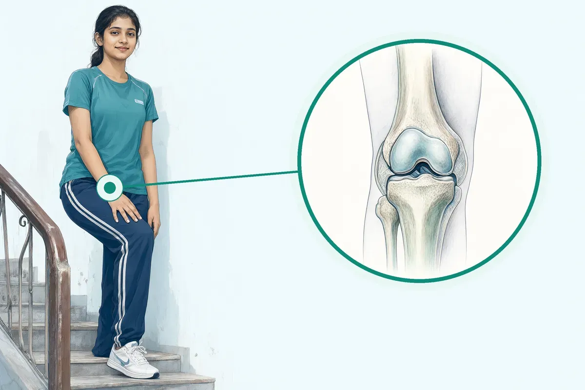
Your kneecap (patella) normally slides up and down inside a vertical groove at the end of your thighbone (femur) when you bend and straighten your leg. A network of tendons and ligaments holds the kneecap in this groove. Patellofemoral disorders happen when the kneecap moves out of this groove or does not track smoothly.
The kneecap can pop completely out of the groove, which is called a dislocation, or it can partly slip out, which is called a subluxation. Most often the kneecap moves to the outer side of the knee. When the kneecap dislocates it locks the knee and pulls the ligaments out of place, often tearing them.
A patella dislocation is different from a knee dislocation, which involves the thighbone and shinbone and is a much more serious injury. Patellofemoral disorders also include maltracking, where the kneecap does not sit centrally in the groove, and chondromalacia, which is softening or wear of the cartilage under the kneecap.
Symptoms and warning signs that need urgent care the same day
- Cannot straighten the knee
- Knee looks deformed
- Cannot bear any weight
- Rapid swelling
- Numbness or tingling in the foot
A dislocated kneecap is painful and stops you from walking. You will not be able to use the leg normally until the joint is corrected. The knee may be locked and unable to straighten or bend, or it may catch and pop when you try to move it.
The joint feels unstable and may buckle when you put weight on it. There is usually a large swelling (effusion) and tenderness along the inner side of the knee. A tense, swollen knee often means there is blood in the joint (haemarthrosis).
If the kneecap pops back into place on its own, the knee will still be sore and swollen from the trauma. You should seek care the same day if you cannot straighten the knee, if the knee looks deformed, if you cannot bear any weight, if the swelling appears rapidly, or if you have numbness or tingling in the foot.
These signs may mean:
- A fracture
- A trapped loose piece of bone or cartilage
- Nerve or blood vessel injury that needs immediate attention
Causes and who is at higher risk
- around 49% Recurrence after second dislocation
- Up to 45% Redislocation after first episode
- up to 95% Cartilage injury in first-time dislocation
A direct blow or a fall can knock the kneecap out of place. A sudden twist of the knee while the foot stays planted on the ground can also cause it. Some people are born with a shallow groove (trochlear dysplasia) or other structural differences that make the kneecap unstable. A family history suggests a genetic link for congenital dislocation.
Risk factors include:
- Being young
- Female
- Having loose ligaments
- A high-riding kneecap (patella alta)
- A tibial tubercle that sits too far to the outer side
A previous dislocation on the other knee raises the risk of recurrence six-fold. After a second dislocation the chance of another episode is much higher, around 49%. Up to 45% of people who have had one dislocation will dislocate again. Articular cartilage injuries are very common, occurring in up to 95% of first-time dislocations.
How patellofemoral disorders are diagnosed: examination and tests
A doctor can usually diagnose a dislocated kneecap by examining your knee and asking about the injury. The physical examination checks for swelling, tenderness on the inner side, and how the kneecap moves. The doctor will also test the ligaments and check the alignment of your leg.
X-rays (anteroposterior, lateral and axial views) are taken first to see the kneecap position and look for a bone fragment. X-rays can miss cartilage injuries, so an MRI is often needed to see the joint surfaces, ligaments and any loose pieces. MRI reliably shows osteochondral injuries in first-time dislocators. If there is a large haemarthrosis, MRI is especially indicated.
A CT scan may be used to measure the tibial tubercle-trochlear groove (TT-TG) distance, which helps plan surgery. MRI can also measure TT-TG distance, but the values may not be interchangeable with CT.
Treatment options without surgery and when surgery is advised
First-time dislocations without a displaced bone fragment are usually treated without surgery. The knee is rested, iced and elevated. A brace or cast in extension may be used for a period, often around six weeks, followed by physiotherapy to regain movement and strengthen the quadriceps. Some studies used shorter periods of immobilisation, but a higher redislocation rate may result.
Pain relief with NSAIDs is common. Most people return to daily activities in six to eight weeks and to sports in three to four months. Surgery is considered when there is a displaced osteochondral fragment large enough to fix (usually with at least 9 mm of bone).
Surgery may also be advised after recurrent dislocations, when the kneecap remains unstable despite rehabilitation, or when there are structural problems such as a very shallow groove or a high TT-TG distance.
Your surgeon will discuss which procedure suits your anatomy, such as:
- MPFL reconstruction
- Tibial tubercle osteotomy
- Lateral release or trochleoplasty
Preparing for patellofemoral surgery
Do
- Tell surgeon about all medicines
- Report fever or illness before surgery
- Fast for at least six hours
- Bring regular medicines and ID
- Pack loose clothing and flat shoes
- Arrange attendant for first night
Before surgery, your surgical team will ask for a set of investigations. These usually include blood tests, a chest X-ray and an ECG to check your fitness for anaesthesia. Imaging of the knee is essential. You will typically have an MRI to look at the cartilage, ligaments and any loose fragments.
A CT scan may also be requested to measure the tibial tubercle-trochlear groove distance, which helps the surgeon plan the procedure.
Tell your surgeon about every medicine you take, including blood thinners, diabetes tablets, insulin and any supplements. Some medicines may need to be stopped a few days before the operation. If you have a fever, cough, skin infection or any new illness in the week before surgery, call the hospital — the date may need to move.
You will be asked to fast for at least six hours before the procedure. This means no food, milk or tea. You may sip a small amount of water with your usual morning tablets if the anaesthetist has agreed. Bring your regular medicines, a list of allergies, your insurance or scheme cards and a photo ID.
Pack loose, comfortable clothing that fits over a bulky knee dressing, and a pair of flat, supportive shoes. An attendant should plan to stay with you for the first night after discharge.
How patellofemoral surgery is done
MPFL reconstruction
- Rebuilds medial ligament with tendon graft
Tibial tubercle osteotomy (Fulkerson)
- Shifts bony attachment medially and forward
Lateral release or lengthening
- Divides tight outer tissues arthroscopically
The operation is performed under spinal or general anaesthesia and usually takes one to two hours. Most patients stay in hospital for one to two nights. The exact procedure depends on the underlying problem — instability, maltracking or cartilage wear — and your surgeon will advise which option suits you.
- MPFL reconstruction: The medial patellofemoral ligament is rebuilt using a tendon graft. This is the common choice for recurrent dislocation where the ligament is torn or stretched.
- Tibial tubercle osteotomy (Fulkerson): The bony attachment of the patellar tendon on the tibia is cut and shifted medially and forward. This offloads worn cartilage and corrects maltracking.
- Lateral release or lengthening: Tight tissues on the outer side of the knee are divided arthroscopically to reduce excessive lateral pressure.
- Trochleoplasty: The groove at the end of the thigh bone is deepened to hold the kneecap more securely. This is reserved for severe trochlear dysplasia.
Sometimes two procedures are combined, for example MPFL reconstruction with a tibial tubercle osteotomy. On the day of surgery you will be admitted to the ward, meet the anaesthetist, and sign the consent form. After the operation you will wake up in the recovery area with a bulky bandage and a knee brace.
Pain relief is given through injections or tablets. The nursing team will check your circulation and sensation in the foot regularly. You will start gentle ankle and toe movements straight away. A physiotherapist will teach you how to use crutches and how much weight you may put on the leg before you go home.
Risks and possible complications of patellofemoral surgery
All operations carry some risk. In a large review of MPFL reconstruction, complications were reported in 26.1 percent of knees (164 complications in 629 knees).
The events ranged from minor to major and included:
- Patellar fracture
- Graft failure
- Persistent instability on examination
- Loss of knee flexion
- Wound problems and pain
Twenty-six patients needed a second operation.
Other known risks include stiffness, weakness and loss of muscle control that can follow prolonged immobilisation. Long-term, the joint may develop osteoarthritis, especially if the cartilage was damaged at the time of the original dislocation. Recurrent dislocation remains possible after surgery, particularly if you return to high-risk sports too early or if the underlying bony anatomy is severely abnormal.
Your surgical team will discuss the risks that apply to your specific procedure and anatomy. They will also explain the steps taken to reduce complications, such as careful surgical technique, early mobilisation and a structured rehabilitation programme.
Recovery and aftercare after patellofemoral surgery
- First few days Ice, elevation, pain tablets, crutches, brace full-time
- Two weeks Stitches or clips removed, physiotherapy starts
- Six to eight weeks Return to daily activities
- Four to six months Expected return to activity at Jaipur Hospital
- More than a year High-level sport may take longer
In the first few days you will manage swelling with ice, elevation and the prescribed pain tablets. Keep the dressing clean and dry. You will walk with crutches, putting only the amount of weight your surgeon allows. The knee brace is usually worn full-time for the first few weeks, then gradually reduced as your quadriceps control returns.
Stitches or clips are removed at about two weeks. Physiotherapy starts early with range-of-motion exercises and static quadriceps work. Over the next weeks you progress to weight-bearing exercises, cycling and strengthening. Most people return to daily activities by six to eight weeks.
Return to sport typically takes three to four months after non-surgical treatment, but after surgery it may take longer — often more than a year for high-level sport. The protocol at Jaipur Hospital expects return to activity in four to six months.
Call the hospital or attend the emergency department if you notice:
- Increasing redness, warmth or discharge from the wound
- A sudden increase in pain not relieved by tablets
- Calf swelling or tenderness
- Fever above 38°C
- If the knee gives way repeatedly
Keep your follow-up appointments so the team can check healing and adjust your rehabilitation plan.
Results of patellofemoral surgery: how well it works and recurrence
- 26.4 points average increase Kujala score improvement
- 4.8 percent Functional failure rate (newer studies)
- 9.5 percent Functional failure rate (older reports)
Studies show that MPFL reconstruction significantly improves patient-reported outcome scores. In a pooled analysis, the Kujala score — a questionnaire about knee function — increased by an average of 26.4 points after surgery. The mean preoperative score ranged from 50 to 70, and postoperative scores reached around 79 in some series. Functional outcomes remain favourable as complication and failure profiles have improved over time.
Redislocation rates after MPFL reconstruction are low in recent studies, with functional failure rates dropping from 9.5 percent in older reports to 4.8 percent in newer ones. However, the likelihood of preventing redislocation appears higher when trochleoplasty is combined with extensor apparatus balancing for severe dysplasia. No single fixation technique or graft choice has been shown to give better functional outcomes than another.
For first-time dislocations, randomised trials have not shown a clear benefit of early surgery over bracing and physiotherapy in terms of final function or activity levels, although some studies report fewer redislocations with surgical repair. After a second dislocation the risk of further episodes rises substantially, and surgery is often considered. Your surgeon will weigh the evidence against your age, activity goals, imaging findings and the number of previous episodes.
Cost and insurance for patellofemoral surgery in Jaipur
The typical price range for patellofemoral realignment surgery at Jaipur Hospital is ₹1,20,000 to ₹3,00,000.
The final amount depends on:
- The procedure performed — MPFL reconstruction, tibial tubercle osteotomy, lateral release, trochleoplasty or a combination
- The type of implants or grafts used
- The length of stay
- Whether you have any additional medical conditions that need care during admission
Ask the admissions desk for a detailed estimate before you are admitted.
Jaipur Hospital is empanelled under RGHS, MAA Yojana and CGHS, and works with a panel of private insurers and TPAs. If you are covered by one of these schemes, bring your scheme card and any pre-authorisation letter. The billing team will help you understand what is covered and what you may need to pay out of pocket. The hospital has an in-house pharmacy and diagnostic laboratory, so medicines and tests during your stay are billed directly.