

Neurosurgery and Neurology · Jaipur Hospital
Minimally Invasive Neurosurgery in Jaipur
You want smaller cuts and faster recovery. We assess your scans and give a clear plan.
- Serving Jaipur since 1991 (35 years)
- NABH accredited
- 24×7 emergency & ICU
- Cashless: RGHS, MAA Yojana, CGHS + 22 insurers
- Operation time
- 2 to 4 hours
- Anaesthesia
- General anaesthesia
- Recovery
- Light activity in 2 to 3 weeks
- Price
- ₹1,50,000 to ₹4,00,000typical range in India; ask us for an estimate
Big cuts, long recovery
Open surgery means large incisions, more pain, and weeks off work
Traditional brain or spine surgery often needs a big opening. That means more muscle damage, longer hospital stays, and a slower return to daily life.
-
Large scar and more pain
Open approaches cut through more muscle and tissue, leaving a bigger wound that hurts longer.
-
Longer hospital stay
You may spend extra nights in the ward or ICU before you can go home.
-
Weeks away from work
Recovery can keep you from your job and family duties for a month or more.
-
Higher infection risk
Bigger wounds and longer operations raise the chance of wound problems.
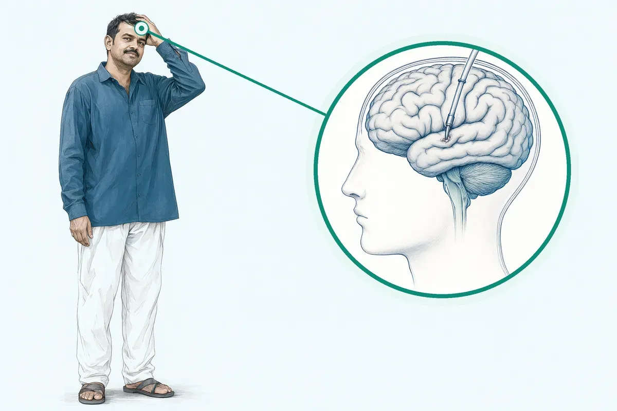
Minimally invasive spine surgery uses smaller incisions and causes less damage to nearby muscles and tissue.
Why Jaipur Hospital
You worry that surgery will keep you bedridden, drain your savings, and leave a scar you have to explain.
Your care plan
Your first steps with us
- 1 Call or visit OPD for a neurosurgery consultation
- 2 Get scans reviewed and tests done in-house
- 3 Discuss keyhole options and receive a cost estimate
- 4 Have surgery and recover with follow-up visits
How the operation is done
The surgeon works through small openings using an endoscope or a tubular retractor. General anaesthesia is used. The procedure usually takes two to four hours.
-
Endoscopic Transsphenoidal
Pituitary tumours reached through the nose without opening the skull.
-
Tubular Microdiscectomy
Slipped disc removed through an 18 mm tube to spare back muscles.
-
Endoscopic Third Ventriculostomy
A new CSF pathway made for hydrocephalus without placing a shunt.
-
Keyhole Craniotomy
A small bone flap gives access to deep brain lesions.
Why patients trust us
- 1991 Serving Jaipur for 35 years
- NABH Accredited hospital
- 1 Neurosurgery and Neurology consultants
- 24×7 Emergency, ICU and ambulance
- 25 Cashless schemes and insurers
What research shows about this treatment
-
Success rate above 60%
ETV for obstructive hydrocephalus succeeds in more than six out of ten patients as first-line treatment.
-
Shorter hospital stay
Endoscopic endonasal approach reduces length of stay compared to traditional transoral surgery.
-
Less blood loss and quicker recovery
Minimally invasive spine techniques show reduced blood loss, shorter hospitalisation, and faster ambulation than open surgery.
If it is left untreated
Untreated brain or spine pressure can cause worsening weakness, vision loss, or bladder and bowel problems. Progressive compression may lead to long-term nerve injury.
Life after treatment
Most people go home in a few days and resume light activity within weeks.
-
Smaller scar
Incisions are a few centimetres long and often hidden in hairlines or natural folds.
-
Less pain
Muscle-sparing approaches usually mean milder discomfort managed with tablets.
-
Faster return to routine
Many patients restart light work and household tasks in two to three weeks.
-
Follow-up imaging
Regular MRI or CT checks every six to twelve months track healing.
Frequently asked questions
Questions patients ask about minimally invasive neurosurgery
Which problems are assessed under minimally invasive neurosurgery?
In short
Minimally invasive neurosurgery at Jaipur Hospital assesses a wide range of brain and spine conditions. These include pituitary tumours, hydrocephalus, intraventricular cysts and tumours, skull base lesions, hypothalamic hamartomas, degenerative spine disease, disc herniation, spinal stenosis, spondylolisthesis and certain craniovertebral junction pathologies. The surgical team reviews imaging and clinical findings to determine if a keyhole or endoscopic approach is suitable.
In detail
Evidence shows neuroendoscopic surgery is now performed for obstructive hydrocephalus, various intraventricular lesions, hypothalamic hamartomas, craniosynostosis, skull base tumours and spinal lesions. The endoscopic endonasal approach addresses pituitary adenoma, Rathke cyst, chordoma, meningioma and craniopharyngioma.
For the spine, minimally invasive techniques treat disc herniation, lumbar spinal stenosis, spondylolisthesis and metastatic spine disease. Transcervical approaches manage odontoid abnormalities and basilar invagination.
The key principle is accessing the target through small corridors with endoscopes or tubular retractors, reducing muscle disruption and blood loss compared to open surgery.
A common belief
Many people think keyhole surgery is only for simple disc problems.
What actually happens
In fact, evidence shows it reaches deep brain tumours, ventricular cysts and complex skull base lesions with similar removal rates to open surgery but less tissue damage.
Your next step at Jaipur Hospital
Your surgeon will review your MRI or CT scans, neurological examination and symptom history at the first visit. If a minimally invasive corridor is feasible, the team discusses the specific technique, typical hospital stay of two to four nights and recovery timeline.
You can ask about the estimated cost range of ₹1,50,000 to ₹4,00,000 and insurance coverage under RGHS, CGHS or private panels.
How will the doctor decide which tests I need?
In short
The neurosurgeon at Jaipur Hospital selects tests based on your symptom pattern, clinical examination findings and the suspected lesion location. Standard investigations include high-resolution MRI of the brain or spine with contrast, CT for bony detail in skull base or spinal cases, and focused blood work including hormonal panels for pituitary lesions. Additional studies like MR angiography, visual field testing or dynamic spine X-rays are ordered only when the surgical plan requires them.
In detail
Evidence guides test selection for each minimally invasive corridor. For endoscopic transsphenoidal surgery, pituitary protocol MRI, endocrine profile and visual fields are essential to map the tumour and assess hormonal function.
For hydrocephalus, MRI with CISS or FIESTA sequences defines ventricular anatomy for endoscopic third ventriculostomy. Spinal cases need MRI with fat suppression and CT for facet joint and pedicle anatomy before tubular microdiscectomy or MIS-TLIF.
Intraoperative navigation and neuromonitoring needs are planned from these preoperative studies. The principle is to obtain only the data that changes surgical decision-making, avoiding redundant radiation or cost.
A common belief
Patients sometimes arrive with a full-body check-up that misses the specific sequences needed for surgical navigation.
What actually happens
The surgeon needs thin-slice MRI with contrast for endoscopic planning, not a routine brain screen. Targeted testing saves time, money and radiation.
Your next step at Jaipur Hospital
The surgeon will list required tests during the consultation. Most imaging can be done at the in-house radiology department. Blood tests use the 24-hour laboratory. If you have recent outside scans, bring the films and reports for comparison. Insurance pre-authorisation may need the test list.
How soon should I expect improvement after minimally invasive neurosurgery?
In short
After minimally invasive neurosurgery at Jaipur Hospital many patients notice a reduction in symptoms such as headache, limb pain or hormonal imbalance within the first few weeks. Light activity is usually possible in two to three weeks. Full recovery varies: some people return to desk work in three to four weeks, while others need several months depending on the procedure, age and general health.
In detail
Studies show that endoscopic transsphenoidal surgery for pituitary tumours often leads to rapid hormonal improvement and headache relief. Tubular microdiscectomy for disc prolapse typically reduces leg or arm pain quickly, though numbness or weakness may take longer to resolve.
Endoscopic third ventriculostomy for hydrocephalus can lower intracranial pressure within days, but shunt independence is confirmed over months. Keyhole craniotomy for deep lesions follows a similar pattern: early wound healing in two weeks, neurological recovery over weeks to months.
Evidence notes that patient compliance and satisfaction are generally better with minimally invasive routes because of less postoperative pain and smaller incisions.
A common belief
A common mistake is expecting instant normalcy.
What actually happens
Nerve healing is slow; numbness or weakness may linger even after the pain is gone. Patience and guided rehab matter.
Your next step at Jaipur Hospital
Plan for at least two weeks of limited activity at home. Arrange an attendant to help with daily tasks for the first few days. Follow the physiotherapy schedule if prescribed.
Keep all follow-up appointments for wound check, imaging and hormone tests if you had pituitary surgery. Contact the neurosurgery team at Jaipur Hospital if new weakness, fever or wound leakage appears. Book your post-operative review before discharge.
How can I decide whether to seek advice about keyhole craniotomy?
In short
Consider seeking advice about keyhole craniotomy at Jaipur Hospital if you have a deep-seated brain lesion — such as a tumour, vascular malformation or abscess — that a neurosurgeon has identified as potentially reachable through a small bone flap. The decision rests on whether the lesion’s location, size and relationship to critical structures allow a safe corridor. Your referring doctor or our neurosurgery OPD can review your MRI and advise if this technique is appropriate.
In detail
Evidence describes keyhole craniotomy as a concept that combines a selected trajectory with a small craniotomy to access intraparenchymal lesions using endoscopic assistance and navigation.
It is typically used for lesions like cavernous angiomas, intraparenchymal haematomas, cerebellar infarctions or brain abscesses where a wide exposure is not needed. The approach aims to minimise brain retraction and skull drilling.
However, total removal may be limited by tumour vascularity, density or instrument constraints. Alternatives include standard craniotomy, stereotactic radiosurgery or conservative monitoring. The surgeon weighs the trade-offs: smaller incision versus limited working angle and possible need for conversion.
A common belief
Patients often assume any brain tumour can be done keyhole.
What actually happens
In reality the corridor must be straight and safe; vascular or large tumours may need a standard opening.
Your next step at Jaipur Hospital
Gather your latest MRI with contrast and any prior reports. Ask your current neurologist for a referral letter. Book a neurosurgery OPD appointment at Jaipur Hospital. Bring an attendant to the consultation.
The team will explain whether a keyhole approach fits your anatomy and outline the typical two to four night stay and price range of ₹1,50,000 to ₹4,00,000. Schedule your review to make an informed choice.
Is minimally invasive neurosurgery always safer than open surgery?
In short
Minimally invasive neurosurgery is not always safer than open surgery. Jaipur Hospital selects the approach based on the lesion type location and your overall health. All surgeries carry risks even minimally invasive procedures. The surgeon may need to convert to an open technique if the anatomy or bleeding makes the keyhole approach unsafe.
In detail
Evidence shows that minimally invasive spine surgery has benefits such as less blood loss less muscle damage shorter hospital stay and quicker recovery. However complications can still occur including infection nerve injury dural tear cerebrospinal fluid leak and the need for a second operation.
In some complex tumours or vascular lesions the limited corridor of a keyhole approach can restrict bleeding control and tumour removal.
Studies report that results in terms of tumour removal symptom relief and complication rates are at least similar to open microsurgical series but not universally better. The surgeon weighs the specific risks of each approach for your condition.
A common belief
Patients often assume smaller incision means zero risk.
What actually happens
The evidence shows complication rates are similar to open surgery for many complex lesions. The choice depends on your specific tumour or spine problem.
Your next step at Jaipur Hospital
Ask your surgeon to explain why a minimally invasive or open approach is recommended for your case. Discuss the specific risks that apply to your lesion and health status. The team at Jaipur Hospital will outline the expected benefits and possible complications.
What changes in strength, sensation, speech, vision, or bladder control need urgent assessment?
In short
Sudden weakness in an arm or leg, new numbness, difficulty speaking or understanding speech, loss of vision or double vision, and loss of bladder or bowel control all need urgent assessment. Jaipur Hospital runs a 24×7 emergency department for these neurological red flags. Do not wait for a routine appointment if these appear.
In detail
These symptoms can signal worsening compression of the brain, spinal cord or nerves from a tumour, haemorrhage, hydrocephalus or postoperative complication such as haematoma or infection. Evidence shows early decompression improves outcomes for conditions like cervicomedullary compression or cauda equina syndrome. The emergency team will perform rapid imaging and involve the neurosurgeon on call to decide on immediate intervention.
A common belief
Patients sometimes hope symptoms will settle, but these signs often mean increasing pressure on the brain or spinal cord.
What actually happens
Delaying can lead to long-term deficit.
Your next step at Jaipur Hospital
If you notice any of these changes, call the emergency number or come directly to the emergency department at Jaipur Hospital. An attendant should accompany you.
Which symptoms should prompt me to see a specialist?
In short
You should see a neurosurgery specialist at Jaipur Hospital if you have persistent headaches with vision changes, unexplained weakness or numbness in limbs, difficulty walking or balance problems, new-onset seizures, bowel or bladder control changes, or progressive neck or back pain radiating to arms or legs. These symptoms may indicate a structural brain or spine lesion that needs imaging and expert assessment.
In detail
Evidence links specific symptom patterns to conditions treated by minimally invasive neurosurgery. Pituitary tumours often cause visual field defects, hormonal changes or headaches. Hydrocephalus presents with gait disturbance, urinary incontinence and cognitive decline. Intraventricular tumours may raise intracranial pressure causing headache, nausea and vomiting.
Spinal cord compression from disc herniation, stenosis or craniovertebral junction abnormalities produces radiating pain, weakness, sensory loss or myelopathy. Hypothalamic hamartomas associate with gelastic seizures and precocious puberty. The surgical team evaluates whether your symptom complex matches a lesion accessible through endoscopic or keyhole corridors.
A common belief
Patients often wait hoping symptoms resolve.
What actually happens
However, progressive neurological deficits from compressive lesions rarely improve without decompression. Early specialist review allows minimally invasive options before irreversible damage occurs.
Your next step at Jaipur Hospital
Keep a symptom diary noting onset, frequency and triggers. Bring any prior MRI, CT or X-ray films and reports to the consultation. The neurosurgeon will perform a focused neurological examination and order targeted imaging if needed. Early evaluation allows timely planning if surgery is indicated.
How will the results change my care plan?
In short
Test results at Jaipur Hospital directly shape whether minimally invasive surgery is offered, which technique is chosen, and the timing of the procedure. Imaging defines lesion size, location, vascularity and relationship to critical structures. Hormonal levels guide pituitary surgery urgency and post-operative replacement needs. Spinal alignment and instability on dynamic X-rays determine if fusion is added to decompression. The surgeon integrates all data to confirm the endoscopic corridor is safe and likely to achieve decompression or resection.
In detail
Evidence shows specific findings alter the surgical approach. Large pituitary tumours with cavernous sinus invasion may need staged or combined approaches. Colloid cysts at the foramen of Monro require assessment of venous anatomy to avoid injury.
Hydrocephalus etiology and patient age influence endoscopic third ventriculostomy success rates — aqueductal stenosis has over 60% success, while congenital hydrocephalus in young children fares worse. Spinal stenosis with instability on flexion-extension views shifts the plan from tubular decompression alone to MIS-TLIF with instrumentation. If imaging shows the lesion is not accessible through a keyhole corridor, open surgery or non-surgical management is recommended.
A common belief
A common belief is that any tumour can be removed endoscopically.
What actually happens
Evidence shows large, vascular or laterally extended lesions may need open surgery. The test results tell the surgeon this upfront, avoiding an unsafe minimally invasive attempt.
Your next step at Jaipur Hospital
After all reports are available, the surgeon reviews them with you, explains the recommended technique, risks, hospital stay and recovery timeline. You can discuss the cost estimate and insurance coverage. If surgery is advised, pre-anaesthesia check-up and consent are completed.
What should I do if symptoms persist or worsen after minimally invasive neurosurgery?
In short
If symptoms such as severe headache, new weakness, fever, wound leakage, or worsening vision occur after minimally invasive neurosurgery at Jaipur Hospital, contact the neurosurgery team immediately or visit the 24×7 emergency department. Do not wait for the next scheduled appointment. Early reporting allows prompt imaging and treatment of complications such as cerebrospinal fluid leak, infection or haematoma.
In detail
Evidence highlights that postoperative cerebrospinal fluid leak occurs in about 11 % of transsphenoidal cases and can lead to meningitis if untreated. Central nervous system infection is reported in about 10 %. Incidental durotomy rates are lower in cervical procedures but still possible.
Conversion to open surgery is rare but may be needed if the minimally invasive corridor does not achieve the goal. Any new neurological deficit, increasing drowsiness, or clear fluid dripping from the nose or wound should be treated as urgent. The hospital’s in-house laboratory and imaging provide rapid MRI, CT and blood tests to confirm the issue.
- Evidence highlights that postoperative cerebrospinal fluid leak occurs in about 11 % of transsphenoidal cases and can lead to meningitis if untreated.
- Central nervous system infection is reported in about 10 %.
A common belief
Some patients think a little leakage or headache is normal and will settle.
What actually happens
It often is not. CSF leak and infection need immediate repair or antibiotics.
Your next step at Jaipur Hospital
Keep the emergency numbers 0141-2551500 and +91-7414-001-001 handy. If you are an out-of-town patient, stay in Jaipur for at least one week after discharge for easy access. Carry your discharge summary and medication list to any hospital you visit. Attend all follow-up visits so the team can catch delayed complications early.
How can I decide whether to seek advice about neuroendoscopic haematoma evacuation?
In short
Consider seeking advice about neuroendoscopic haematoma evacuation at Jaipur Hospital if you or a relative has a spontaneous intracerebral haemorrhage or post‑traumatic haematoma that imaging shows may be accessible through a small endoscopic port. The neurosurgeon evaluates haematoma size, location, depth, and the patient's neurological grade to decide if minimally invasive evacuation offers advantage over medical management or open surgery.
In detail
Endoscopic evacuation uses a narrow sheath and irrigation‑suction to remove clot with minimal brain disruption. Evidence indicates it can reduce mass effect and lower peri‑haematomal oedema.
It is typically considered for supratentorial haemorrhages 30–50 ml in volume, with a Glasgow Coma Scale above a certain threshold, and where the clot is not too diffuse. Risks include re‑bleeding, infection, and incomplete evacuation if the clot is organised.
Alternatives include conservative care with close monitoring, stereotactic aspiration, or open craniotomy for larger or superficial clots. The surgeon will discuss the expected benefit for your specific scan findings.
A common belief
Not every clot needs surgery, and not every clot can be removed endoscopically.
What actually happens
Size, location, and patient condition all matter — the goal is to reduce pressure without causing new injury.
Your next step at Jaipur Hospital
If a recent CT or MRI shows a haemorrhage and the treating doctor suggests surgical opinion, request a referral to the Neurosurgery OPD. Bring all imaging on a CD or film.
The surgeon will explain the procedure, typical two to four night stay, and recovery expectations. Timely evaluation is important — early evacuation may improve outcomes in selected cases.
FAQ library
Questions and answers about minimally invasive neurosurgery
When you contact us
Get your assessment and plan
Contact Jaipur Hospital for a neurosurgery OPD consultation, scan review, plain-language options, a cost estimate, cashless insurance help, and follow-up care.
- A consultation with a neurosurgeon in the OPD
- Your scans and reports reviewed, with tests done in-house if you need them (laboratory, X-ray and imaging)
- Your options explained in plain words, including whether you need surgery at all
- A cost estimate before you decide
- Help with cashless treatment under RGHS, MAA Yojana, CGHS and empanelled insurers and TPAs
- Follow-up visits after your treatment
Overview
Minimally Invasive Neurosurgery: what to know
What minimally invasive neurosurgery treats and how the condition develops
Minimally invasive neurosurgery covers keyhole brain and spine surgery using endoscopes and tubular retractors.
The conditions treated include:
- Pituitary tumours
- Rathke cysts
- Chordomas
- Meningiomas
- Nasopharyngeal carcinoma
- Squamous cell carcinoma at the skull base
It also addresses:
- Intraventricular tumours
- Hypothalamic hamartomas
- Craniosynostosis
- Degenerative spine disease
- Rare subtypes of hydrocephalus
In the spine, common problems are slipped discs, spinal stenosis, spondylolisthesis, and metastatic spine disease.
These conditions develop in different ways. Pituitary adenomas grow from the pituitary gland and can press on nearby nerves or cause hormone changes. Hydrocephalus builds up when cerebrospinal fluid (CSF) cannot drain properly, raising pressure inside the skull. Degenerative spine disease comes from wear and tear on discs and joints, leading to nerve compression.
Hypothalamic hamartomas are congenital malformations linked to gelastic seizures, early puberty, and cognitive problems. In children, craniosynostosis happens when skull sutures fuse too early, and the optimal age for endoscopic correction is around three months.
Symptoms and warning signs that need urgent care the same day
- Sudden severe headache
- New weakness or numbness
- Loss of bladder or bowel control
- Difficulty speaking or understanding
- Sudden vision loss
- Worsening drowsiness or confusion
Symptoms depend on the location and type of problem. Brain and skull base lesions may cause headaches, vision changes, hormone imbalances, or seizures. Hydrocephalus often presents with headache, nausea, vomiting, and drowsiness. Spine problems typically produce back or neck pain that radiates to the arms or legs, numbness, weakness, or difficulty walking. Retro-odontoid pseudotumours can cause severe neck pain, cervicomedullary compression, and myelopathy.
Warning signs that need same-day medical attention include sudden severe headache, new weakness or numbness in the face, arm, or leg, loss of bladder or bowel control, difficulty speaking or understanding speech, sudden vision loss, and worsening drowsiness or confusion. If you notice any of these, go to the emergency department immediately.
Causes and who is at higher risk
- Older age
- Linked to higher CSF leak rate after transsphenoidal surgery
- Non-ideal BMI
- Associated with higher postoperative CSF leak and meningitis rates
- Combined open and endoscopic approach
- Raises complication risk; staging procedures can reduce it
- Longer procedure duration
- Primary contributor to postoperative complications
Causes vary by condition. Pituitary tumours and many skull base tumours arise without a clear cause, though some genetic syndromes increase risk. Hydrocephalus can be congenital or follow bleeding, infection, or tumour. Degenerative spine disease relates to ageing, repeated strain, and sometimes injury. Hypothalamic hamartomas and craniosynostosis are congenital.
Higher risk factors include increasing age, which is linked to a higher rate of CSF leak after transsphenoidal surgery. A non-ideal body mass index (BMI) is associated with higher postoperative CSF leak and meningitis rates. Combining open and endoscopic approaches in one sitting raises complication risk, which can be reduced by staging procedures.
Large colloid cysts increase the risk of venous injury at the foramen of Monro. For endoscopic endonasal approaches, risk factors include infection, dural tears, vertebral artery injury, CSF leak, and tracheotomy. Longer procedure duration is the primary contributor to postoperative complications.
How the condition is diagnosed: examination and tests
Diagnosis starts with a detailed history and neurological examination.
Your surgeon will check:
- Strength
- Sensation
- Reflexes
- Coordination
- Cranial nerves
Imaging is the mainstay. MRI of the brain or spine with contrast shows tumours, cysts, hydrocephalus, and nerve compression. CT scans give detail of bone anatomy, especially for skull base and spine planning. For pituitary lesions, hormone blood tests are essential. In hydrocephalus, MRI helps identify the cause and guides whether endoscopic third ventriculostomy (ETV) is suitable.
Endoscopic tumour biopsy is a well-established method for intraventricular brain tumours, with a high diagnostic yield above 90% and low risk below 3.5%.
Germ cell tumours, infiltrative hypothalamic or optic pathway gliomas, and Langerhans cell histiocytosis are addressable with endoscopic biopsy.:
- Germ cell tumours
- Infiltrative hypothalamic or optic pathway gliomas
- Langerhans cell histiocytosis
For spine problems, imaging pinpoints the exact level of compression, which helps the surgeon decide if a minimally invasive corridor is feasible.
Treatment options without surgery and when surgery is advised
Non-surgical options are tried first for many conditions. For degenerative spine disease, this includes medications, physiotherapy, activity modification, and sometimes epidural steroid injections. For small pituitary tumours that are not pressing on nerves or causing hormone excess, observation with regular MRI and hormone checks may be appropriate. Hydrocephalus from infection or bleeding may need temporary CSF diversion before definitive surgery. In children with craniosynostosis, helmet therapy alone is not enough; surgery is required.
Surgery is usually advised when non-surgical treatment has not relieved symptoms, when there is progressive neurological deficit, or when a tumour needs removal or biopsy. For obstructive hydrocephalus due to aqueductal stenosis, ETV is the first-line approach with a success rate above 60%. ETV is also suitable for postoperative hydrocephalus as an alternative to shunt insertion.
For hypothalamic hamartomas, all patients except those with precocious puberty require surgical treatment. In metastatic spine disease, mini-open and percutaneous procedures are considered for shorter recovery and reduced morbidity. Your surgeon will review whether your spine or brain lesion is accessible through small surgical corridors and whether the exact cause of symptoms can be pinpointed.
Preparing for minimally invasive neurosurgery
Do
- Regular medicines
- List of allergies
- Recent test reports
- Comfortable loose clothing
- An attendant
Before minimally invasive neurosurgery, you will meet your surgeon for a consultation. They will perform a physical exam and order imaging tests such as MRI or CT to plan the procedure. Your surgeon will give you specific instructions to follow before surgery.
You should discuss all medicines you take, including blood thinners, supplements, and any hormone replacements, because some may need to be stopped or adjusted. Fasting is required before general anaesthesia; typically you must not eat or drink for six to eight hours before the operation. On the day of surgery, an anaesthesiologist will give you anaesthesia.
For brain and spine procedures at Jaipur Hospital, general anaesthesia is used so you will be asleep and feel no pain. Bring your regular medicines, a list of allergies, any recent test reports, comfortable loose clothing, and an attendant who can stay with you. The hospital has a 24×7 emergency department, in-house pharmacy, and diagnostic laboratory if any last-minute tests are needed.
How minimally invasive neurosurgery is done
- Endoscopic Transsphenoidal Through the nose for pituitary and skull base lesions
- Tubular Microdiscectomy Slipped disc removal through an 18 mm tube
- Endoscopic Third Ventriculostomy New CSF pathway for hydrocephalus without a shunt
- Keyhole Craniotomy Small bone flap for deep brain lesions with navigation
Minimally invasive neurosurgery uses small incisions and specialised tools to reach the brain or spine with less damage to muscles and soft tissue. Your surgeon will advise which technique suits you based on the location and nature of the problem. The main options are:
- Endoscopic Transsphenoidal: for pituitary tumours and skull base lesions through the nose, without opening the skull.
- Tubular Microdiscectomy: for slipped disc removal through an 18 mm tube.
- Endoscopic Third Ventriculostomy (ETV): for hydrocephalus to create a new CSF pathway without a shunt.
- Keyhole Craniotomy: a small bone flap for deep brain lesions, often with navigation assistance.
On the day of surgery, you will be taken to a modular operation theatre. The procedure typically takes 2 to 4 hours. Your surgeon will explain how long they expect your surgery to take. Intraoperative imaging or navigation may be used for precision. After the operation, you will move to a recovery area while the anaesthetic wears off.
Risks and possible complications of minimally invasive neurosurgery
- about 11% CSF leak in endoscopic endonasal surgery
- about 10% CNS infection in endoscopic endonasal surgery
- 1.4% Incidental durotomy cervical spine
- 3.8% Incidental durotomy thoracic spine
- 7.8% Incidental durotomy lumbar spine
All surgeries have risks, even minimally invasive procedures. Common risks of minimally invasive spine surgery include infection, bleeding, nerve injury, dural tear, and cerebrospinal fluid (CSF) leak. In studies of endoscopic endonasal surgery, CSF leak occurred in about 11% of patients and central nervous system infection in about 10%.
Injury to the internal carotid artery, especially the cavernous segment, is a serious but rare complication; it occurs more often on the left side. Higher body mass index and increasing age are associated with higher rates of postoperative CSF leak and meningitis. Combining endoscopic and open approaches in the same sitting raises the risk of CSF leak and infection.
For transoral approaches, postoperative infection rates are higher and may need debridement and flap coverage. In minimally invasive spine surgery, incidental durotomy rates are lower in the cervical region (1.4%) compared to thoracic (3.8%) and lumbar (7.8%). On rare occasions, the surgery may not go as planned and you might need a second surgery or conversion to an open procedure.
You will have one or more small scars; the location depends on the procedure. Contact your surgeon if you notice worsening pain, fever, wound discharge, new weakness, or severe headache after surgery. Go to the emergency room if you have difficulty breathing, sudden severe headache, or loss of consciousness.
Recovery and aftercare after minimally invasive neurosurgery
- Same day to 2–4 nights Hospital stay at Jaipur Hospital
- 2–3 weeks Light activity usually possible
- Several weeks Avoid strenuous activities and heavy lifting
- Weeks to months Full recovery depending on cause, complexity, age and health
- Every 6–12 months Follow-up MRI or CT scans
After surgery, you will stay in the recovery area until the anaesthetic wears off. If you had general anaesthesia, you may feel groggy. Some patients go home the same day, while others stay overnight or for a couple of days depending on the procedure. The typical hospital stay at Jaipur Hospital is 2 to 4 nights.
You will have some discomfort, but it is usually less intense than after open surgery. Pain relievers will be given to manage pain. Before discharge, you will receive instructions on incision care, safe activities, and follow-up appointments. Light activity is usually possible in 2 to 3 weeks. Avoid strenuous activities and heavy lifting for several weeks.
You may need help at home for a few days to weeks. Your doctor may recommend physiotherapy to regain strength and speed up recovery. Full recovery can take weeks to months depending on the cause of symptoms, the complexity of the procedure, your age, and general health.
Keep all follow-up appointments so your care team can check your progress and answer any concerns. Follow-up MRI or CT scans are typically done every 6 to 12 months.
Results of minimally invasive neurosurgery in studies
- above 60% ETV success in aqueductal stenosis
- 5.3% (percutaneous) vs 14.8% (open) Complication rate for traumatic spinal fractures
- lower in minimally invasive group Medical complications
In studies, minimally invasive neurosurgery achieves outcomes at least similar to open microsurgical series for mass removal, symptom relief, and complication rates. The endoscopic approach gives a superior close-up view and wider working angle. For obstructive hydrocephalus, ETV is a first-line treatment with success rates above 60% in aqueductal stenosis, and equally effective for tectal plate lesions.
Success rates are better in older children and adolescents (over 70%) but lower in very young children with congenital hydrocephalus or myelomeningocele. Results depend on the cause of hydrocephalus and patient age. For hypothalamic hamartomas, endoscopic disconnection appears safer and more effective than other modalities. Endoscopic endonasal excision of pituitary adenomas and craniopharyngiomas shows encouraging results with low morbidity.
Both endoscopic and microscopic approaches are equally effective for nonfunctioning adenomas without large suprasellar or lateral extensions. For larger tumours, the endoscopic approach may improve extent of resection and reduce postoperative complications. In minimally invasive spine surgery, meta-analyses show reduced blood loss, shorter time to ambulation, and shorter hospital stay compared to open surgery.
Medical complications are lower in the minimally invasive group. For traumatic spinal fractures, percutaneous approaches reduce complication rates from 14.8% (open) to 5.3%. However, operative time is not significantly different, and long-term clinical outcomes such as fusion rates are similar between minimally invasive and open techniques.
The success rate varies based on the technique used and is measured by reduction of symptoms like pain by managing the underlying cause. Patient compliance is reported to be better with endoscopic approaches.
Cost and insurance for minimally invasive neurosurgery in Jaipur
The typical price range for minimally invasive neurosurgery in India is ₹1,50,000 to ₹4,00,000. The final cost depends on the type of procedure, the implants or endoscopic tools needed, the length of hospital stay, ICU requirements, and any complications that may arise.
Hospital bills usually include surgery fees (surgeon, assistant, anaesthesia team), operation theatre costs, hospital stay (room rent, ICU, nursing), pre-operative investigations (MRI, CT, blood tests), anaesthesia and medicines, post-operative care (follow-up imaging, hormone tests, rehabilitation), and consumables (implants, disposables, endoscopic tools).
Long-term medications, especially hormone replacements after pituitary surgery, and rehabilitation therapy (speech, physiotherapy, occupational therapy) add to the overall cost. Follow-up MRI or CT scans every 6 to 12 months are also an expense to plan for.
Most comprehensive health insurance plans in India cover brain surgeries, including endoscopic procedures. Jaipur Hospital is empaneled with RGHS, MAA Yojana, CGHS, and a panel of private insurers and TPAs. Check whether the hospital is empaneled with your insurer. Understand what portion of the cost is reimbursed, as room rent caps often limit coverage.
Get pre-authorisation before admission to avoid delays. Keep all bills and medical records for smooth claim processing. Ask whether post-operative medications and follow-up scans are covered. Direct billing arrangements with insurers can reduce upfront payments. For patients without insurance or with partial coverage, ask the hospital for a written cost estimate before admission.