

Plastic Surgery · Jaipur Hospital
Skin Lesions and Skin Cancer in Jaipur
Worried about a changing mole or lump? Get a clear diagnosis and a simple treatment plan.
- Serving Jaipur since 1991 (35 years)
- NABH accredited
- 24×7 emergency & ICU
- Cashless: RGHS, MAA Yojana, CGHS + 22 insurers
- Procedure time
- 15 to 60 minutes
- Anaesthesia
- Local anaesthesia
- Recovery
- Suture removal at 7 to 14 days, normal activity next day
- Price
- ₹5,000 to ₹50,000typical range in India; ask us for an estimate
A spot that will not heal
You noticed a change and do not know what it means
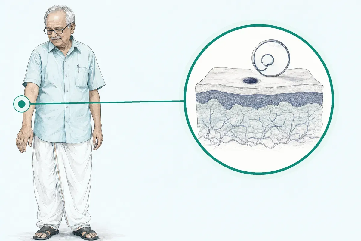
A mole that changes colour, a lump that grows, a sore that will not heal. You wonder if it is serious. You worry about cancer, about scars, about time off work and the cost of treatment.
-
Changing mole or new growth
A mole that changes shape, colour or size, or a new spot that looks different from the rest.
-
Lump under the skin
A cyst or lipoma you can feel, sometimes painful, sometimes just visible.
-
Sore that will not heal
A scab or ulcer that bleeds, crusts over and returns after weeks.
-
Cosmetic concern
A benign lesion on the face or neck that makes you self-conscious.
-
Fear of cancer
You have read the warning signs and want a proper check, not guesswork.
Melanomas can develop within a mole you have had for a long time or show up as a new mole.
Why Jaipur Hospital
You want to know quickly whether it is dangerous, without long waits or hidden costs.
Your care plan
Your first steps with us
- 1 Call or visit OPD for a surgeon consultation
- 2 Get examined, dermoscopy and biopsy if needed
- 3 Review the plan and cost estimate with us
- 4 Have daycare surgery and return for suture removal
How the operation is done
Most procedures take 15 to 60 minutes under local anaesthesia. You go home the same day. Stitches come out at 7 to 14 days.
-
Excision Biopsy
Complete removal with a margin of healthy skin. The specimen goes for pathology. Stitches close the wound.
-
Shave or Punch Biopsy
A sample is taken for diagnosis first. A razor or circular blade removes the top layers. No stitches usually needed.
-
Mohs Micrographic Surgery
Layer-by-layer removal with immediate margin check under a microscope. Used for high-risk cancers on the face.
-
Cryotherapy or Electrocautery
Freezing or heat destroys the lesion. Used for benign growths and very superficial cancers. No specimen for pathology.
Why patients trust us
- 1991 Serving Jaipur for 35 years
- NABH Accredited hospital
- 1 Plastic Surgery consultants
- 24×7 Emergency, ICU and ambulance
- 25 Cashless schemes and insurers
What research shows about this treatment
-
2 to 3 weeks
Healing after mole removal usually takes two to three weeks, during which daily activities can continue.
-
more likely to come back
Moles removed by shave excision are somewhat more likely to recur than those removed by surgical excision.
-
low-risk procedure
Office-based mole removal is a low-risk procedure, though all surgeries carry some risk of bleeding, infection or scar.
If it is left untreated
If a cancerous lesion is left untreated, it can grow deeper and spread to other parts of the body. Early removal with clear margins reduces that risk.
Life after treatment
Most people return to normal routine the next day. The wound heals in a few weeks and the specimen tells you if further steps are needed.
-
Back to work next day
You can usually drive home and resume daily activities immediately after the procedure.
-
Clear answer from biopsy
The lab report confirms whether the cells were normal or if you need more treatment.
-
Small scar that fades
A fine line scar is typical. It usually fades over months with sun protection.
-
Peace of mind
Knowing the diagnosis lets you stop worrying and plan any next steps calmly.
Frequently asked questions
Questions patients ask about skin lesions and skin cancer
Which problems are assessed under skin lesions and skin cancer?
In short
Jaipur Hospital assesses a wide range of skin lesions and skin cancers through its surgical team. This includes common benign growths such as moles, cysts and lipomas as well as suspicious lesions that may represent skin cancer. The service covers diagnosis and surgical removal for all these conditions.
In detail
The surgical team evaluates moles that appear atypical or have changed in size, shape or colour, as these can sometimes develop into melanoma. Cysts and lipomas are assessed when they cause discomfort, become infected or raise cosmetic concerns.
For lesions that look suspicious, a biopsy is performed to confirm whether cancer cells are present. The evidence shows that most moles are harmless but any lesion that changes, bleeds or itches should be examined.
The team uses dermoscopy to examine skin patterns closely before deciding on the appropriate biopsy technique such as shave, punch or excisional biopsy.
A common belief
Many people think only dark or raised moles need checking.
What actually happens
In fact, any lesion that changes colour, shape, size or starts to itch or bleed should be examined. Even flesh-coloured cysts or soft lipomas can need removal if they grow or cause discomfort. Self-checks at home often miss early changes that a dermoscopy can detect.
Your next step at Jaipur Hospital
If you notice a new or changing skin lesion, you can book a consultation at Jaipur Hospital for a thorough assessment. The surgeon will examine the lesion, discuss whether a biopsy or removal is needed and explain the typical daycare procedure. Suture removal usually occurs at 7 to 14 days and normal activity can resume the next day.
How will the doctor decide which tests I need?
In short
At Jaipur Hospital, the surgeon decides which tests you need after a clinical examination and dermoscopy of the skin lesion. The choice depends on the lesion's appearance, size, location and whether it looks suspicious for cancer. A biopsy is the main test to confirm the diagnosis before planning definitive treatment.
In detail
The evidence explains that the dermatologist or surgeon selects the biopsy technique based on the type of lesion and the amount of tissue the pathologist needs. For flat or slightly raised lesions, a shave or punch biopsy may be used to sample tissue.
For deeper or suspicious lesions, an excisional biopsy removes the entire lesion with a margin of normal skin for full assessment. Dermoscopy helps visualise patterns not visible to the naked eye, guiding the decision.
If the lesion appears benign such as a typical lipoma or cyst, the surgeon may proceed directly to excision without a separate biopsy. The in-house diagnostic laboratory processes the tissue sample for histopathological analysis.
A common belief
Patients often expect a blood test or scan for skin lesions.
What actually happens
In reality, the only reliable test for most skin growths is a tissue biopsy examined under a microscope. Imaging like ultrasound or MRI is rarely needed unless the surgeon suspects deep spread. Dermoscopy at the bedside gives immediate information that guides the biopsy type better than any scan for surface lesions.
Your next step at Jaipur Hospital
During your consultation at Jaipur Hospital, the surgeon will examine your lesion, possibly take clinical photographs for monitoring and explain which biopsy or excision is appropriate. The procedure is usually done as daycare under local anaesthesia taking 15 to 60 minutes. Results from the laboratory typically guide the next steps within a few days.
How soon should I expect improvement after the chosen treatment?
In short
At Jaipur Hospital, the wound typically heals in two to three weeks. You may have stinging or burning for a few days. Normal activity can resume the next day. Suture removal is at 7 to 14 days. The final scar appearance improves over several months.
In detail
After excision or shave removal, the area is kept clean, moist and covered. Daily dressing changes are advised. Stinging or burning is common for the first few days. Healing time depends on your health and the size of the lesion.
Most people return to work or school immediately. The scar matures over months; applying sunscreen after healing helps minimise pigmentation. If the lesion was removed for cancer, the pathology result confirms whether margins are clear. Clear margins mean the local treatment is complete. Recurrence is possible but less likely with excision than with shave removal. Follow-up is scheduled based on the diagnosis.
A common belief
Some people expect the scar to vanish quickly.
What actually happens
Scars take months to mature and may remain visible, though they fade with time and sun protection.
Your next step at Jaipur Hospital
Keep the wound moist with petroleum jelly and change the dressing daily. Avoid strenuous activity that stretches the wound for the first week. Attend suture removal at 7 to 14 days. Apply sunscreen to the healed area. Contact the surgeon if you notice increasing pain, redness, swelling or discharge.
What do the type, grade, and stage of a skin lesion mean?
In short
Type refers to the kind of cells involved, such as basal cell carcinoma, squamous cell carcinoma, or melanoma. Grade describes how abnormal the cells look under the microscope. Stage indicates how far the cancer has spread, based on size, depth, and whether lymph nodes or distant organs are involved. At Jaipur Hospital, the pathology report provides these details after biopsy.
In detail
Evidence shows that melanoma can develop in a long-standing mole or appear as a new lesion. Basal cell and squamous cell carcinomas are more common and rarely spread.
The pathologist examines the biopsy sample to determine the cell type (type), the degree of cellular abnormality (grade), and features like Breslow thickness or ulceration that help assign a stage. Staging may require additional imaging or sentinel lymph node biopsy for thicker melanomas.
These factors guide the treatment plan: low-risk lesions may need only excision with clear margins, while higher-stage disease may need wider excision, lymph node assessment, or systemic therapy. Your surgeon will explain what the specific type, grade, and stage mean for your situation.
A common belief
People often confuse grade and stage.
What actually happens
Grade is about cell appearance; stage is about spread. Both matter for treatment choices.
Your next step at Jaipur Hospital
After biopsy, the pathology report is usually available within a few days. Your surgeon will review the results with you and outline the recommended treatment. If further staging tests are needed, they will be arranged promptly.
How can I decide whether to seek advice about mole and nevus excision?
In short
You should seek advice about mole excision at Jaipur Hospital if a mole changes colour, shape or size, becomes itchy, bleeds, or looks different from your other moles. The ABCDE rule helps: Asymmetry, Border irregularity, Colour variation, Diameter over 6 mm, Evolving. Cosmetic concern is also a valid reason. A dermatologist can assess and advise if removal is needed.
In detail
Evidence shows most moles are harmless and do not need treatment. Typical moles are round, flat or slightly raised, even in colour and stable over time. However, melanomas can develop within a long-standing mole or appear as a new mole.
Atypical moles look unusual but are not melanoma. Your healthcare provider might recommend removing a mole if it looks suspicious. Some people also have moles removed for cosmetic reasons. A dermatologist is a medical doctor who specialises in diagnosing and treating skin diseases.
They may use a dermoscope to examine the mole closely. If a mole looks concerning, a biopsy is done so the cells can be examined under a microscope. The biopsy technique depends on the mole type and the tissue needed for diagnosis. Do not attempt at-home removal because you cannot biopsy the tissue and may miss cancer.
A common belief
Many people ignore changing moles or try home remedies.
What actually happens
Home removal products cannot biopsy the tissue, so cancer can be missed. Even lasers or cautery are not usually recommended for moles because they destroy the sample needed for diagnosis. Only a medical professional can safely remove and test the mole.
Your next step at Jaipur Hospital
Check your moles monthly in good light. Note any change in size, shape, colour, or symptoms like itching or bleeding. If you notice any ABCDE feature or a mole that simply bothers you, book a consultation. The surgeon will examine you, discuss options and explain the procedure.
How will appearance, function, and realistic expectations be discussed for skin lesion surgery?
In short
At Jaipur Hospital, your surgeon will examine the lesion, explain the planned technique — excision, shave, punch, or Mohs — and describe the expected scar size and location. You will discuss the likely cosmetic outcome, any functional impact, and the healing timeline before you consent.
In detail
Evidence shows that surgical excision with sutures leaves a linear scar that fades over months. Shave excision may leave a round, slightly depressed mark. Mohs surgery aims to minimise the defect on the face. Most patients are happy with the appearance after healing.
The surgeon will explain that a biopsy scar is usually worth the peace of mind of knowing if a mole is melanoma. You will be advised on wound care — keeping it moist, covered, and protected from sun — to optimise the scar.
If reconstruction with a flap or graft is needed for a larger defect, the surgeon will show you what to expect. No promise of a specific cosmetic result can be made, but the team aims for the most suitable functional and aesthetic outcome.
A common belief
Some patients expect invisible scars.
What actually happens
In reality, every cut leaves a mark. The goal is a fine line that fades. Shave excision can leave a small round mark. Mohs on the face preserves tissue but still leaves a scar. Good aftercare makes the biggest difference.
Your next step at Jaipur Hospital
Ask your surgeon about the expected scar length, suture removal at 7 to 14 days, and when you can resume normal activity. Follow the aftercare instructions closely.
Which symptoms should prompt me to see a specialist?
In short
You should see a specialist at Jaipur Hospital if a mole or skin lesion changes in size, shape or colour, becomes itchy, bleeds or develops an irregular border. New moles appearing in adulthood or lesions that look different from your other moles also warrant assessment. These changes can indicate atypical cells or early skin cancer.
In detail
The evidence highlights that most moles are harmless but any change in appearance should be evaluated. The ABCDE rule helps identify concerning features: asymmetry, border irregularity, colour variation, diameter larger than 6 millimetres and evolution over time.
Symptoms such as persistent itching, tenderness, bleeding without injury or a sore that does not heal within a few weeks are also significant. Cysts and lipomas that grow rapidly, become painful, red or warm may be infected and need surgical review. The surgical team uses a dermoscope to examine these features closely before recommending a biopsy or removal.
A common belief
People often wait until a mole hurts or bleeds heavily.
What actually happens
However, early skin cancers rarely cause pain. A flat, dark spot that slowly widens or a pink pearly bump that never heals can be more serious than a sore mole. Self-diagnosis using photos online often misses subtle changes that dermoscopy reveals.
Your next step at Jaipur Hospital
If you notice any of these symptoms, schedule a consultation at Jaipur Hospital. The surgeon will perform a detailed skin examination, possibly using dermoscopy, and advise whether a biopsy or excision is needed. The procedure typically takes 15 to 60 minutes under local anaesthesia with suture removal at 7 to 14 days.
How will the results change my care plan?
In short
The biopsy results from Jaipur Hospital determine whether further treatment is needed. If the cells are normal, no further surgery is usually required. If abnormal or cancerous cells are found, the surgeon will discuss the next steps which may include wider excision, additional imaging or referral to oncology depending on the type and stage of skin cancer.
In detail
The evidence states that when a mole is removed and the cells look normal and healthy, you will not need any further treatment. If the cells appear very abnormal or cancerous, the dermatologist helps figure out the next steps.
For basal cell or squamous cell carcinoma, a wider excision with clear margins is often curative. For melanoma, the thickness of the tumour guides whether sentinel lymph node biopsy, imaging or systemic therapy is needed.
The in-house laboratory provides histopathology reports that the surgical team reviews to tailor the plan. The evidence also notes that if the surgeon achieves good margins during removal, the procedure may serve as curative treatment for skin cancer that has not spread.
A common belief
Patients often assume a biopsy result means either everything is fine or they need chemotherapy.
What actually happens
In reality, many skin cancers like basal cell carcinoma are fully treated by a second wider excision alone. Only deeper melanomas or advanced squamous cell cancers need scans, lymph node checks or systemic therapy. The histopathology report tells the surgeon exactly how much more tissue to remove or whether other specialists should be involved.
Your next step at Jaipur Hospital
After your biopsy or excision at Jaipur Hospital, the surgical team will contact you with the histopathology results usually within a few days. If further surgery is needed, it will be scheduled promptly as a daycare procedure. For complex cases, a multidisciplinary discussion may be arranged.
What should I do if symptoms persist or worsen after skin lesion removal?
In short
If you notice increasing pain, spreading redness, swelling, bleeding that does not stop with pressure, pus, fever, or the wound opening after skin lesion removal at Jaipur Hospital, contact your surgeon promptly. These signs may indicate infection or delayed healing that needs medical review.
In detail
Evidence shows that mole removal is a low-risk procedure, but complications such as infection, bleeding, or wound breakdown can occur. You may feel stinging or burning for a few days, which is normal.
Keeping the area clean, moist with petroleum jelly, and covered with a fresh dressing each day reduces the chance of infection. If the wound becomes more painful, hot, or discharges pus, or if you develop a temperature above 38°C, these are warning signs.
Recurrence of a lesion after shave excision is also possible, so any new growth at the site should be shown to your surgeon.
A common belief
Many people think mild redness means infection, but some pinkness is normal for a few days.
What actually happens
The real warning signs are worsening pain, heat, pus, or fever.
Your next step at Jaipur Hospital
Continue daily wound care as instructed and avoid soaking the area until sutures are removed at 7 to 14 days. Most people return to normal activity the next day. If any concerning symptom appears, call the hospital or visit the OPD for a quick check.
Has the skin condition spread, and which tests can assess that?
In short
Whether a skin lesion has spread depends on its type, depth, and high-risk features seen on biopsy. At Jaipur Hospital, the surgical team uses clinical examination, sentinel lymph node biopsy for eligible melanomas, and imaging such as ultrasound, CT, or PET-CT when staging is required.
In detail
Evidence indicates that for thin melanomas (under 0.8 mm without ulceration) the risk of spread is low, so routine imaging is not recommended. For thicker melanomas or those with ulceration, sentinel node biopsy is offered to check the first draining lymph node.
For high-risk squamous cell carcinoma, imaging of the regional nodes and sometimes chest or abdomen may be advised. Basal cell carcinoma rarely spreads, so staging tests are seldom needed.
The decision is individualised: your surgeon reviews the pathology, examines the lymph node basins, and orders only the tests that change management. Unnecessary scans expose you to radiation and cost without benefit.
A common belief
Many patients request full-body scans immediately.
What actually happens
Guidelines recommend against routine imaging for early lesions because false positives cause anxiety and further invasive tests. The right test at the right time is safer and more useful.
Your next step at Jaipur Hospital
After your biopsy results are available, the surgeon will discuss whether staging investigations are indicated. If sentinel node biopsy or imaging is recommended, it will be scheduled promptly.
FAQ library
Questions and answers about skin lesions and skin cancer
When you contact us
Get your lesion assessed this week
Book an OPD consultation with our plastic surgeon, get a cost estimate and see if you qualify for cashless treatment under your scheme.
- A consultation with a plastic surgeon in the OPD
- Your scans and reports reviewed, with tests done in-house if you need them (laboratory, X-ray and imaging)
- Your options explained in plain words, including whether you need surgery at all
- A cost estimate before you decide
- Help with cashless treatment under RGHS, MAA Yojana, CGHS and empanelled insurers and TPAs
- Follow-up visits after your treatment
Overview
Skin Lesions and Skin Cancer: what to know
What skin lesions and skin cancer are
- Mole
- Round or oval, even colour, stable size
- Cyst
- Benign lump under the skin
- Lipoma
- Benign fatty lump under the skin
- Atypical mole
- Looks unusual but is not melanoma
A skin lesion is any area of skin that looks different from the skin around it.
It can be:
- A mole
- A cyst
- A lipoma
- A patch that has changed colour or texture
Most moles are harmless. They often appear during childhood or the teenage years and can be flat or raised, ranging in colour from your natural skin tone to pink, brown or black. People with darker hair or skin tend to have darker moles.
Typical moles are:
- Round or oval
- Even in colour
- Stay much the same shape and size over time
It is normal to have about 10 to 40 moles by the time you are an adult.
Sometimes a mole can become cancerous or precancerous. Melanoma can develop inside a mole you have had for a long time or appear as a new mole. Atypical moles look a little unusual but are not melanoma. Other common lesions include cysts and lipomas, which are benign lumps under the skin. A dermatologist is a doctor who specialises in diagnosing and treating diseases of the skin.
Symptoms and warning signs that need urgent care
- Changes colour or shape
- Becomes itchy or bleeds
- Not round or oval
Most moles do not need treatment. You should see your healthcare provider if you have any concerns about the appearance of a mole, or if it changes colour or shape, becomes itchy or bleeds. Contact your healthcare provider if your mole is itchy, bleeds, is not round or oval, or you notice changes in its appearance.
You should check your moles regularly and note any changes. A mole that looks suspicious may be recommended for removal so it can be tested. Some people also choose to have a mole removed for cosmetic reasons.
Causes and who is at higher risk
- Fair skin
- Higher risk of abnormal moles
- History of sunburn
- Increases risk of skin cancer
- Family history of skin cancer
- Genetic risk factor
- Many or atypical moles
- Increases chance of change over time
Moles form when pigment cells in the skin grow in clusters. Sun exposure and genetics play a part in how many moles you develop and how they look. People with fair skin, a history of sunburn or a family history of skin cancer may have a higher risk of developing abnormal moles or skin cancer.
Having many moles or atypical moles also increases the chance that one could change over time. Your healthcare provider can help you understand your personal risk during a skin check.
How skin lesions and skin cancer are diagnosed
- Shave biopsy
- Blade removes surface sample
- Punch biopsy
- Circular tool removes deeper sample
- Excisional biopsy
- Entire lesion cut out with margin
Your healthcare provider will inspect your skin and may take pictures of your moles to compare later. They may use a tool called a dermoscope to get a closer look at the mole and decide how to remove it. If a mole looks concerning, a biopsy is done so the cells can be examined under a microscope.
The dermatologist selects the biopsy technique based on the type of mole and the amount of tissue the pathologist needs.
Common methods include:
- Shave biopsy
- Punch biopsy
- Excisional biopsy
These are minor surgical procedures. Any time a mole is removed, it is important to check that the cells are normal. Having a mole biopsied is a simple, low-risk procedure.
Treatment options without surgery and when surgery is advised
Dermatologists may use cryotherapy or electrocautery for some benign lesions and superficial skin cancers. These methods are not usually recommended for moles because they do not provide a tissue sample for biopsy. Laser removal is also not suitable for moles, as a biopsy might still show the mole is cancerous.
Shave excision and surgical excision with sutures are two commonly used methods to remove a mole. Moles removed by shave excision are somewhat more likely to come back than those removed by surgical excision. Moles are more likely to recur after nonsurgical treatments. Sometimes benign moles that come back after nonsurgical removal can show features of skin cancer.
Your surgeon will advise which technique suits you. Surgery is advised when a lesion looks suspicious, when a diagnosis is needed or when a lesion causes discomfort or cosmetic concern.
Preparing for skin lesion and skin cancer surgery
Do
- Tell surgeon about blood-thinning medicines
- Tell team about allergies
- Bring medicine list and insurance card
- Wear loose clothing
Your surgeon will review your medical history and any medicines you take. Blood-thinning medicines such as aspirin, clopidogrel or warfarin may need to be stopped a few days before the procedure, but only change or stop a medicine after speaking with the doctor who prescribed it. Tell the team about any allergies, especially to local anaesthetic or adhesive dressings.
Routine blood tests are not usually required for small lesions removed under local anaesthesia, but your surgeon will advise if any tests are needed. You do not need to fast for local anaesthesia; you can eat and drink normally on the day of surgery. Wear loose, comfortable clothing that does not rub against the lesion site.
Bring a list of your current medicines, your insurance or scheme card if you have one, and an attendant to help you home afterwards. If the lesion is on your face or scalp, avoid make-up or hair products on the day of surgery.
How skin lesion and skin cancer surgery is done
- Numb the area with local anaesthetic Injection or numbing cream
- Clean and mark the lesion Surgeon prepares the site
- Remove lesion using chosen technique Excision, shave, punch, Mohs, cryotherapy or electrocautery
- Send specimen to pathology lab Cells examined under microscope
The procedure is performed in a daycare operating theatre and usually takes 15 to 60 minutes. You will be awake and the area will be numbed with local anaesthetic given as an injection; sometimes a topical numbing cream is applied first.
The surgeon cleans the skin, marks the lesion and removes it using one of the following techniques, chosen according to the type, size and site of the lesion and whether cancer is suspected:
- Excision biopsy: the entire lesion is cut out with a margin of normal skin and the wound is closed with stitches. This provides a full specimen for diagnosis and, if margins are clear, can be curative for skin cancer.
- Shave or punch biopsy: a blade or a circular punch tool removes a sample of the lesion for diagnosis before definitive treatment is planned. Shave biopsy does not assess deep margins and cannot distinguish between types of skin cancer.
- Mohs micrographic surgery: the lesion is removed layer by layer and each layer is examined under a microscope immediately. This technique is used for high-risk cancers on the face or other cosmetically sensitive areas to spare healthy tissue while ensuring clear margins.
- Cryotherapy or electrocautery: liquid nitrogen or an electric current destroys the lesion. These methods are used for benign lesions and some superficial cancers, but no tissue is available for biopsy and recurrence is more likely.
After removal, the specimen is sent to the pathology laboratory. The wound is dressed and you can go home the same day.
Risks and possible complications of skin lesion and skin cancer surgery
Mole and lesion removal under local anaesthesia is a low-risk procedure.
Common minor issues include:
- A small scar
- Temporary stinging or burning for a few days
- Mild swelling
- Bruising
- A small amount of bleeding during or after the procedure
Infection at the wound site is uncommon but possible; keeping the wound clean and covered reduces this risk. Recurrence of the lesion can happen, especially after shave excision, cryotherapy or electrocautery, because these methods may leave deeper cells behind.
In studies of advanced melanoma treatments, grade 3 or 4 treatment-related adverse events occurred in 13.1% of patients receiving nivolumab and 35.1% receiving standard therapy, but these figures relate to systemic immunotherapy, not to local surgical removal. If a removed mole grows back after healing, contact your surgeon for review. Your surgeon will discuss the specific risks for your lesion type and chosen technique before you consent.
Recovery and aftercare after skin lesion and skin cancer surgery
- Same day Go home after procedure
- Next day Return to normal daily activities
- 7 to 14 days Stitches removed if used
- Two to three weeks Healing complete
You can usually drive yourself home and return to normal daily activities the next day. The wound should be kept clean, moist and covered; apply petroleum jelly or the ointment provided and change the dressing daily or as instructed. Stitches, if used, are typically removed at 7 to 14 days.
Healing takes about two to three weeks depending on the size and site of the lesion and your general health. You may have a little soreness during this time but it should not stop you from doing your usual activities. After the wound has healed, apply sunscreen to the area when it is exposed to the sun.
Contact the hospital or your surgeon if you notice:
- Increasing redness
- Swelling
- Warmth
- Pus
- Worsening pain
- Bleeding that does not stop with pressure
- If you develop a fever
Also call if the lesion appears to be growing back after it has healed.
Results of skin lesion and skin cancer surgery
Most moles and benign lesions removed by surgical excision are permanently removed. When excision biopsy achieves clear margins, it can serve as curative treatment for skin cancer that has not spread. If pathology shows normal cells, no further treatment is needed.
If abnormal or cancerous cells are found, your surgeon will discuss the next steps, which may include wider excision or referral to a cancer specialist. A biopsy scar is usually worth the peace of mind of knowing whether an abnormal mole is melanoma.
Shave excision provides a tissue sample for analysis and patients are generally pleased with the cosmetic result after healing, but it is not useful for assessing deep tumour margins and recurrence is somewhat more likely than with full excision.
In studies of advanced melanoma, combination immunotherapy with nivolumab plus ipilimumab showed a median overall survival of more than 60 months and a 5-year overall survival of 52%, compared with 26% for ipilimumab alone, but these results apply to advanced metastatic disease treated with systemic therapy, not to local surgical removal of early skin cancers.
Your surgeon will explain what the pathology result means for you and whether any further treatment or monitoring is recommended.
Cost and insurance for skin lesion and skin cancer surgery in Jaipur
The typical price range for surgical removal of a skin lesion or skin cancer in India is ₹5,000 to ₹50,000.
The final cost depends on:
- Size and site of the lesion
- Technique used (excision biopsy, shave or punch biopsy, Mohs surgery, cryotherapy or electrocautery)
- Whether a wider margin or reconstruction is needed
- Pathology charges
Jaipur Hospital is a NABH-accredited multispeciality hospital established in February 1991. We accept patients under RGHS, MAA Yojana, CGHS and a panel of private insurers and TPAs. The hospital has modular operation theatres, an in-house diagnostic laboratory and imaging, a 24-hour laboratory, X-ray and ECG, and an in-house pharmacy. As a daycare procedure, no overnight stay is required. Please ask us for an estimate based on your specific case.