

Sports Injury · Jaipur Hospital
Knee Cartilage and Meniscus in Jaipur
Knee pain, locking or giving way? Get a clear assessment and a simple plan at Jaipur Hospital.
- Serving Jaipur since 1991 (35 years)
- NABH accredited
- 24×7 emergency & ICU
- Cashless: RGHS, MAA Yojana, CGHS + 22 insurers
- Operation time
- 45 to 90 minutes
- Anaesthesia
- Spinal or General anaesthesia
- Recovery
- Full weight bearing in 4 to 6 weeks
- Price
- ₹80,000 to ₹2,50,000typical range in India; ask us for an estimate
Knee pain stops you
Your knee hurts, locks or gives way when you walk or climb stairs
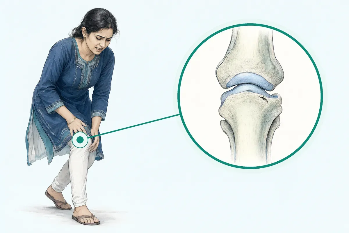
A meniscus tear or cartilage damage can make the knee catch, swell or feel unstable. Everyday tasks like walking, squatting or sitting cross-legged become difficult. Pain may wake you at night. You may worry about long-term damage or needing a major operation later.
-
Pain and swelling
Knee aches after activity, swells by evening, and may hurt at rest or at night.
-
Locking or catching
The knee gets stuck in one position or clicks painfully when you move it.
-
Giving way
The knee buckles without warning on stairs or uneven ground.
-
Limited movement
You cannot fully bend or straighten the knee, making sitting cross-legged hard.
-
Fear of worsening
You worry the damage will spread and lead to early arthritis.
The meniscus is important for load distribution, shock absorption and stability of the knee joint.
Why Jaipur Hospital
You want to walk without pain, keep working and avoid a knee replacement later, but you are not sure if surgery is necessary or what it involves.
Your care plan
Your first steps with us
- 1 Book an OPD consultation with a sports injury specialist
- 2 Get scans and tests done in-house if needed
- 3 Review options and a cost estimate with your surgeon
- 4 Have arthroscopic surgery and start guided rehab
How the operation is done
Surgery is usually done under spinal or general anaesthesia through two or three small keyhole cuts. The procedure takes 45 to 90 minutes. You typically go home the same day or stay one night.
-
Meniscal Repair
All-inside or inside-out sutures stitch the torn edges together so the meniscus can heal. Used for peripheral tears with good blood supply.
-
Partial Meniscectomy
The unstable flap is trimmed away while preserving as much healthy meniscus as possible.
-
Microfracture / Nanofracture
Tiny holes are made in the bone to release marrow cells that form repair tissue over a small cartilage defect.
-
OATS / Mosaicplasty
Healthy bone and cartilage plugs are moved from a non-weight-bearing area to fill a larger defect.
Why patients trust us
- 1991 Serving Jaipur for 35 years
- NABH Accredited hospital
- 1 Sports Injury consultants
- 24×7 Emergency, ICU and ambulance
- 25 Cashless schemes and insurers
What research shows about this treatment
-
Full recovery in a few weeks to a few months
Meniscectomy recovery is usually around six weeks; meniscus repair takes longer because the meniscus must heal inside the knee.
-
No difference in pain or function at 12 months
In a randomised trial, arthroscopic partial meniscectomy gave similar improvement in knee scores as sham surgery at one year.
-
Infection in about 1 in 100 cases
A serious knee infection occurred in one participant after partial meniscectomy in a large trial; no serious events were seen in the sham group.
If it is left untreated
Untreated unstable tears can accelerate cartilage wear and increase the risk of osteoarthritis in that knee. Meniscectomy removes tissue that protects the joint, which may raise long-term arthritis risk.
Life after treatment
Most people return to daily activities and light sport after guided rehabilitation. Recovery speed depends on the procedure done.
-
Walk without support
Most people walk without crutches within a few weeks after meniscectomy; longer after repair.
-
Return to work
Desk work often resumes in one to two weeks; heavy labour takes longer.
-
Sport and recreation
Light activity usually returns in a month or two; contact sports may need several months.
-
Confidence in the knee
With rehab, most people regain trust in the knee for stairs, squatting and uneven ground.
Frequently asked questions
Questions patients ask about knee cartilage and meniscus
Which problems are assessed under knee cartilage and meniscus?
In short
At Jaipur Hospital, the knee cartilage and meniscus service assesses meniscus tears, cartilage defects, and related knee joint problems. The surgical team evaluates traumatic tears, degenerative lesions, and focal cartilage damage. Your surgeon will check for mechanical symptoms such as locking or giving way, and for signs of early osteoarthritis.
In detail
The meniscus acts as a shock absorber and stabiliser in the knee. Tears can be traumatic from twisting injuries or degenerative from ageing tissue. Cartilage defects may be small focal lesions or wider wear.
Evidence shows degenerative meniscal lesions are often the first sign of osteoarthritis and are frequently seen on MRI in middle-aged people. The team uses clinical examination and imaging to grade the tear pattern, location, and stability.
Longitudinal vertical tears in the peripheral zone have good healing potential, while complex or radial tears may be stable. The decision to operate depends on symptom severity, tear type, and whether nonoperative care has been tried.
A common belief
Many people think every tear needs surgery, but evidence shows degenerative tears often improve with physiotherapy first.
What actually happens
The team checks tear type, location, and your symptoms before recommending any procedure.
Your next step at Jaipur Hospital
You can book an OPD consultation for a clinical assessment and MRI review. The surgeon will explain the findings and discuss whether physiotherapy, activity modification, or surgery is appropriate. Most patients are seen within a few days.
How will the doctor decide which tests I need?
In short
At Jaipur Hospital, your surgeon decides on tests after taking a detailed history and examining your knee. The clinical assessment checks for joint line tenderness, effusion, range of motion, and mechanical symptoms such as locking or giving way. Based on these findings, the surgeon orders the most relevant imaging.
In detail
Plain X-rays are usually the first test to assess bone alignment, joint space narrowing, and osteophytes. Weight-bearing views help grade osteoarthritis using the Kellgren-Lawrence scale.
MRI is requested when soft tissue detail is needed, such as confirming a meniscus tear pattern, evaluating cartilage defects, or checking ligament integrity. Evidence shows degenerative meniscal lesions are frequently seen on MRI in asymptomatic middle-aged people, so imaging is always interpreted alongside your symptoms.
The surgeon may also use the Western Ontario Meniscal Evaluation Tool or Lysholm score to quantify your functional limitation. Blood tests are rarely needed unless infection or inflammatory arthritis is suspected.
A common belief
MRI often shows tears in people with no pain.
What actually happens
The surgeon matches the scan findings to your actual symptoms and examination, not the image alone.
Your next step at Jaipur Hospital
Tests are usually done in-house on the same day or within 24 hours. The surgeon reviews the results with you at the next visit and explains how they change the treatment plan.
How soon should I expect improvement after knee cartilage or meniscus surgery?
In short
At Jaipur Hospital, most patients notice reduced swelling and less pain within the first two weeks after arthroscopic meniscus or cartilage surgery. Full weight bearing usually begins at four to six weeks, and functional improvement continues for several months depending on the exact procedure performed.
In detail
Evidence from randomised trials shows that knee scores such as Lysholm and WOMET improve steadily over the first twelve months after both meniscal repair and partial meniscectomy.
In studies, mean improvements of twenty to thirty points were common at twelve months, with no significant difference between surgical and sham groups for degenerative tears.
Recovery is faster after meniscectomy — around six weeks — while meniscal repair or microfracture needs three months or more for the tissue to heal. Your surgeon will guide the timeline based on the technique used and your knee's condition.
A common belief
Many people expect a quick fix, but studies show improvement is gradual.
What actually happens
Degenerative tears often do just as well with exercise alone, so surgery is not always faster.
Your next step at Jaipur Hospital
Plan for two weeks of limited activity, then progressive physiotherapy. Most people return to desk work within one to two weeks and to light duties by four weeks. Heavy labour or sport may need three to six months.
Can a meniscus or cartilage injury heal with rehabilitation alone, or is surgery worth considering?
In short
Many degenerative meniscus tears and small cartilage defects improve with a structured exercise programme, and surgery is not always needed. At Jaipur Hospital, your surgeon will compare the expected outcomes of physiotherapy versus arthroscopic repair, debridement or microfracture based on your tear type, symptoms and activity goals.
In detail
High-quality trials show that for degenerative tears without mechanical locking, exercise therapy yields similar pain relief and function to arthroscopic partial meniscectomy at twelve and twenty-four months.
In one study, thirty percent of patients randomised to exercise alone later chose surgery, meaning seventy percent avoided it.
However, traumatic vertical tears in the vascular zone, bucket-handle tears causing locking, and unstable flaps often do better with early repair to preserve meniscal function and protect cartilage. Microfracture or OATS are considered for focal cartilage defects that fail non-operative care. The decision balances symptom severity, tear pattern, age, and osteoarthritis grade.
A common belief
Patients often assume surgery is the only fix.
What actually happens
Studies show exercise gives equal results for many degenerative tears, and seventy percent avoid surgery altogether.
Your next step at Jaipur Hospital
Start with a supervised neuromuscular and strengthening programme for twelve weeks. If mechanical symptoms persist or function plateaus, review with your surgeon.
How can I decide whether to seek advice about meniscus repair?
In short
You should consider seeking advice about meniscus repair at Jaipur Hospital if you have a traumatic tear, a longitudinal vertical tear in the outer third of the meniscus, or mechanical symptoms such as locking or giving way. Repair is usually the first option to preserve meniscal function, especially in younger patients.
In detail
Evidence indicates that longitudinal vertical tears in the vascularised peripheral zone have high healing rates and protect cartilage long term. Repair shows better clinical outcomes and less osteoarthritis progression compared with meniscectomy.
Nonoperative treatment is first-line for degenerative tears, but traumatic root tears, ramp lesions with ACL injury, and radial tears are strong indications for repair. Your surgeon will assess tear pattern, location, and knee stability on MRI and examination before recommending repair.
A common belief
Many assume all tears are trimmed.
What actually happens
Evidence shows repair gives better long-term cartilage protection and less arthritis than meniscectomy, but degenerative tears often do well with exercise first.
Your next step at Jaipur Hospital
If your knee locks, gives way, or has persistent swelling after a twisting injury, book a consultation at Jaipur Hospital. The surgeon will review imaging, explain whether your tear pattern suits repair, and discuss the typical four-to-six-week weight-bearing timeline. Early advice helps preserve the meniscus and joint health.
Can I continue any training safely while I recover from knee cartilage or meniscus surgery?
In short
Yes, you can continue modified training during recovery from knee cartilage or meniscus surgery at Jaipur Hospital, but the type and intensity depend on the procedure. After meniscectomy, stationary cycling and upper-body work often start within days. After repair or microfracture, protected weight-bearing limits lower-body loading for several weeks.
In detail
Evidence supports early motion and cardiovascular maintenance to prevent deconditioning. The physiotherapy team prescribes a graduated programme: range-of-motion exercises first, then non-weight-bearing strengthening (straight-leg raises, hip work), progressing to partial then full weight-bearing as healing allows.
Swimming with a pull buoy, upper-body ergometry, and core work are usually safe throughout. Running, jumping, and cutting are restricted until strength and hop symmetry criteria are met, typically three to six months after repair or microfracture.
A common belief
Many athletes either stop completely or push too hard.
What actually happens
Both extremes delay recovery — inactivity causes muscle loss, while early loading can damage the healing tissue.
Your next step at Jaipur Hospital
Your surgeon and physiotherapist will give you a written weekly plan showing what is allowed at each stage. Follow the load progression closely — doing too much too soon risks graft failure or repair disruption.
Which symptoms should prompt me to see a specialist?
In short
You should see a specialist at Jaipur Hospital if you have knee pain, swelling, stiffness, catching, locking, or a feeling that the knee gives way. These symptoms may indicate a meniscus tear or cartilage injury. Persistent symptoms that limit daily activities or disturb sleep also warrant an orthopaedic opinion.
In detail
Common symptoms include pain along the joint line, swelling that appears hours or days after activity, and mechanical sensations such as clicking, catching, or the knee locking in one position. Instability or buckling suggests a displaced tear.
Degenerative tears often cause gradual onset pain and stiffness, especially after sitting. Evidence indicates that mechanical symptoms like locking are linked to unstable tear patterns such as bucket-handle or flap tears.
The surgeon correlates your symptom pattern with clinical tests and imaging to determine if the meniscus or cartilage is the source.
A common belief
People often wait until the knee locks completely.
What actually happens
Early evaluation helps distinguish mechanical tears that may need repair from degenerative changes that respond to physiotherapy.
Your next step at Jaipur Hospital
Make an OPD appointment if symptoms last more than a few weeks, worsen, or interfere with walking, stairs, or sleep. Bring any previous X-rays or MRI scans.
The surgeon will examine your knee, may order an MRI, and then explain whether physiotherapy, activity modification, or a procedure is needed.
How will the results change my care plan?
In short
At Jaipur Hospital, test results directly shape your care plan. MRI findings on tear type, location, and cartilage health determine whether nonoperative treatment, repair, debridement, or cartilage restoration is offered. The surgeon matches the lesion characteristics to your age, activity level, and symptom severity.
In detail
Evidence shows that degenerative tears without mechanical symptoms often do well with exercise therapy alone. Peripheral vertical tears in the vascular zone are candidates for repair to preserve meniscal function. Unstable flap tears in the avascular zone may need partial meniscectomy.
Cartilage defect size and depth guide choice between microfracture for small lesions and osteochondral transfer for larger ones. Osteoarthritis grade on X-ray influences whether meniscus surgery is likely to help.
The surgeon explains which findings are relevant to your symptoms and which are incidental, then outlines options with expected recovery timelines.
A common belief
A tear on MRI does not automatically mean surgery.
What actually happens
Many degenerative tears improve with exercise. The surgeon treats your symptoms, not just the scan, and explains which findings matter for your plan.
Your next step at Jaipur Hospital
After reviewing your MRI and X-rays, the surgeon discusses the treatment pathway: observation with physiotherapy, a daycare arthroscopic procedure, or a more involved cartilage restoration. You receive a clear explanation of why each option fits your findings.
What should I do if symptoms persist or worsen after knee cartilage or meniscus surgery?
In short
If pain, swelling, locking or giving way continue or get worse after your arthroscopic procedure at Jaipur Hospital, contact your surgical team promptly. Early review helps identify infection, re-tear, stiffness or other issues that may need medication, further physiotherapy or occasionally a second arthroscopy.
In detail
Evidence indicates that adverse events such as knee infection occur in a small minority of cases, and reoperation rates are higher after meniscal repair than after partial meniscectomy. Persistent mechanical symptoms like locking or giving way may signal a residual unstable fragment or new tear.
Stiffness can develop if early mobilisation is inadequate. Your surgeon will examine the knee, review imaging if needed, and decide whether to adjust rehabilitation, prescribe anti-inflammatory medication, or plan further intervention. Do not ignore new fever, increasing redness, or sudden inability to bear weight.
A common belief
Many patients expect a straight-line recovery.
What actually happens
In reality, a small percentage develop infection, re-tear or stiffness, and repair procedures have higher reoperation rates than meniscectomy.
Your next step at Jaipur Hospital
Keep a symptom diary noting activity level, swelling timing and pain scores. Attend all scheduled follow-ups at two weeks, six weeks and three months. If concerns arise between visits, call the hospital's orthopaedic department or visit the 24×7 emergency department.
Does the scan show a partial tear, complete tear, or wear-related change in my meniscus or cartilage?
In short
MRI at Jaipur Hospital classifies meniscus tears as partial or complete and describes their pattern — longitudinal, radial, horizontal, bucket-handle or complex. It also grades cartilage defects from surface softening to full-thickness loss and notes any bone oedema or osteoarthritis signs.
In detail
Evidence indicates that longitudinal vertical tears in the outer third have good healing potential with repair, while radial, horizontal and complex patterns are often stable and may not need surgery. Degenerative tears are frequent in middle-aged knees and commonly coexist with early osteoarthritis.
Cartilage defects are sized and located; focal lesions under one to two centimetres may respond to microfracture, whereas larger areas may need osteochondral transfer.
Your surgeon correlates imaging findings with your examination to decide whether the tear is unstable and symptomatic or an incidental age-related change.
A common belief
Patients often assume any tear on MRI needs surgery.
What actually happens
Many degenerative tears are asymptomatic incidental findings, and treatment depends on matching symptoms to the imaging, not the scan alone.
Your next step at Jaipur Hospital
Bring your MRI images and report to the orthopaedic OPD. The surgeon will explain the exact tear morphology, cartilage grade and treatment implications in plain language.
FAQ library
Questions and answers about knee cartilage and meniscus
When you contact us
Get a clear plan for your knee
Contact Jaipur Hospital for an OPD consultation, in-house scans, a plain-word explanation of your options, a cost estimate and help with cashless schemes.
- A consultation with a sports injury specialist in the OPD
- Your scans and reports reviewed, with tests done in-house if you need them (laboratory, X-ray and imaging)
- Your options explained in plain words, including whether you need surgery at all
- A cost estimate before you decide
- Help with cashless treatment under RGHS, MAA Yojana, CGHS and empanelled insurers and TPAs
- Follow-up visits after your treatment
Overview
Knee Cartilage and Meniscus: what to know
What a meniscus tear and cartilage damage are
- Meniscus
- C-shaped cartilage between thigh and shin bone
- Two per knee
- Each knee has two menisci
- Function
- Shock absorbers that spread body weight
- Cartilage defect
- Worn or damaged smooth surface on bone ends
The meniscus is a C-shaped piece of cartilage that sits between your thigh bone and shin bone. Each knee has two menisci. They act like shock absorbers, spreading your body weight across the joint and keeping the knee stable when you move. A meniscus tear happens when this cartilage rips.
Cartilage defects are areas where the smooth surface lining the ends of the bones has worn away or been damaged.
Most meniscal tears are degenerative. This means the tissue weakens with age and can tear during normal daily movements, not just during sport. A tear can also happen suddenly when you twist your knee while your foot stays planted, which is common in sports. When the meniscus is torn or removed, the knee loses some of its shock absorption.
This increases the load on the joint surface and can speed up the wear that leads to osteoarthritis. A tear at the root of the meniscus is especially important because it stops the meniscus from working properly and can accelerate cartilage damage.
Symptoms of a meniscus tear and when to seek urgent care
- Knee locked and cannot straighten it
- Knee hot, very swollen, and feverish
Common symptoms include:
- Knee pain or tenderness
- Stiffness
- Swelling around the knee
The swelling may not appear for a few hours or even days after the injury. You may feel the knee give way or buckle, or it may lock or get stuck in one position. Some people hear or feel a popping sensation when the tear happens.
Contact a doctor the same day if you have very bad knee pain after a fall or injury.
You should also seek prompt medical help if the knee is locked and you cannot straighten it, if you cannot put any weight on the leg, or if the knee becomes hot, very swollen, and you feel feverish, as these can suggest an infection or a serious injury that needs quick assessment.
Causes and who is at higher risk
- Over 45 with osteoarthritis
- Early changes on imaging increase risk
- Pivoting sport athletes
- Sudden twists during play cause tears
- Repeated deep kneeling or squatting
- Work movements stress the meniscus
- Previous ACL tear
- Ligament injury makes meniscus tears more likely
Degenerative tears are linked to ageing of the tissue and are often the first sign of osteoarthritis in the knee. They are frequently seen on MRI scans in middle-aged and older adults, even in people who do not have symptoms. Traumatic tears usually happen during a sudden twist or deep squat, often while playing sport.
People at higher risk include:
- Those over 45 with early osteoarthritis changes on imaging
- Athletes who play pivoting sports
- Anyone whose work involves repeated deep kneeling or squatting
A previous injury to the knee ligaments, such as an ACL tear, can also make meniscus tears more likely, especially ramp lesions at the back of the meniscus.
How a meniscus tear or cartilage defect is diagnosed
Your doctor will ask about your symptoms and examine your knee. They will check for tenderness along the joint line, swelling, and how well the knee moves. Specific tests, where the doctor bends and rotates your knee, help suggest a meniscus tear.
An MRI scan is the main imaging test used to confirm the diagnosis. It shows the tear pattern, its location, and whether the tear is stable or unstable. Unstable patterns include longitudinal, bucket-handle, and flap tears, while radial, horizontal, and complex tears are often stable. X-rays may be taken to look for osteoarthritis or other bone changes.
Before any surgery, you will be asked about all medicines and supplements you take, and you may need to stop some of them temporarily.
Non-surgical treatment and when surgery is advised
Non-operative treatment is the first-line choice for degenerative meniscal lesions. This usually means a structured physiotherapy programme to improve strength, movement, and control of the knee. In studies, exercise therapy improved function and activity levels in patients with degenerative tears, whether or not they later had surgery. Pain relief medicines and activity modification can also help.
Surgery may be advised if you have a serious (high-grade) tear or if non-surgical treatments have not controlled your symptoms. Repair is preferred when the tear is in the outer, vascular zone of the meniscus, because it has a better chance of healing. Longitudinal vertical tears in the peripheral third are considered the standard indication for repair.
Other clear indications for repair include:
- Posterior ramp lesions with an ACL tear
- Traumatic root tears
- Radial lesions
Horizontal cleavage tears in young athletes are also repaired to avoid removing the meniscus. Partial meniscectomy (trimming the torn part) is usually reserved for cases where repair is not possible or has failed, or when there are considerable mechanical symptoms like locking.
Your surgeon will discuss which option fits your tear pattern, age, activity level, and the condition of the rest of your knee.
Preparing for knee cartilage and meniscus surgery
Do
- Attend pre-operative OPD check-up Surgeon reviews MRI, X-rays and symptoms
- Tell surgeon all medicines and supplements Include blood thinners, diabetes tablets, herbal items
- Bring crutches, medicines and an attendant Attendant stays with you and takes you home
- Carry insurance card and pre-authorisation letter For RGHS, MAA Yojana, CGHS or private TPA
Before surgery you will attend a pre-operative check-up at the OPD. The surgeon will review your MRI and any X-rays and ask about symptoms such as locking, giving way or swelling. You will have routine blood tests, an ECG and a chest X-ray if needed for your age or medical history.
Tell your surgeon about every medicine you take, including blood thinners, diabetes tablets, herbal supplements and painkillers bought from a chemist. Some medicines may need to be stopped a few days before the operation. You will meet the anaesthetist who will explain spinal or general anaesthesia and ask about allergies or previous problems with anaesthesia.
You must not eat or drink for about 12 hours before the scheduled time. Wear loose, comfortable clothes that can fit over a bulky knee bandage. Bring your regular medicines, a pair of crutches if you already have them, and an attendant who can stay with you and take you home.
The hospital provides a gown, basic toiletries and meals for the attendant. If you are covered under RGHS, MAA Yojana, CGHS or a private TPA, carry your card and any pre-authorisation letter.
How knee cartilage and meniscus surgery is done
- Small cuts made around the knee Two or three portals for camera and instruments
- Joint filled with sterile fluid Allows clear view of meniscus and cartilage
- Surgeon chooses technique for your tear Repair, meniscectomy, microfracture or OATS
The operation is performed through two or three small cuts (portals) around the knee. A camera (arthroscope) and fine instruments are inserted. The joint is filled with sterile fluid so the surgeon can see the meniscus and the cartilage surface clearly.
Your surgeon will choose the technique that matches the tear pattern, its location and the condition of the surrounding cartilage. The main options are:
- Meniscal repair: stitching the tear with all-inside or inside-out sutures. This is usually offered for fresh, vertical tears in the outer third of the meniscus where blood supply is good.
- Partial meniscectomy: trimming away the unstable flap or shredded part while keeping as much healthy meniscus as possible. This is chosen when the tear is in the inner avascular zone or is complex and cannot heal.
- Microfracture or nanofracture: making tiny holes in the exposed bone to stimulate a repair tissue for small, focal cartilage defects.
- OATS or mosaicplasty: moving a plug of bone and cartilage from a non-weight-bearing part of your own knee to fill a larger defect.
The surgeon will also check the ligaments and the rest of the joint during the same sitting. The procedure usually takes 45 to 90 minutes. At the end, the fluid is drained, the portals are closed with stitches or adhesive strips and a bandage is applied.
You wake up in the recovery room where nurses monitor your pain, sensation and leg movements until the anaesthesia wears off.
Risks and possible complications of knee cartilage and meniscus surgery
- Serious complications are uncommon
- Infection, blood clots, nerve injury are rare
- Removing meniscus increases osteoarthritis risk
- Higher chance of X-ray changes at five years
- Repair preserves shock absorption
- Linked to less degenerative change over time
Knee arthroscopy is a common and generally safe operation, but every surgery carries some risk.
The most frequent problems are:
- Temporary swelling
- Stiffness
- Mild pain around the portals
These usually settle with rest, ice, elevation and the prescribed pain medicines.
In studies, serious complications are uncommon. Infection in the joint (septic arthritis) was reported in about 1 participant in a large trial. Blood clots in the leg veins (deep vein thrombosis) or lung (pulmonary embolism) are rare after this type of short procedure, especially when you start moving the ankle and walking early. Nerve or blood vessel injury around the knee is very rare.
A specific long-term consideration is that removing meniscus tissue increases the load on the knee cartilage. Research shows that partial meniscectomy is associated with a higher chance of osteoarthritis changes on X-ray at five years compared with non-operative care. Repairing the meniscus, when possible, preserves its shock-absorbing function and is linked to less degenerative change over time. Re-operation rates are somewhat higher after repair than after meniscectomy, usually because the tear does not heal or re-tears.
Your surgeon will discuss the risks that apply to your tear type, age, activity level and any existing arthritis. Call the hospital or your surgeon if you develop increasing redness, warmth, fever, calf pain, sudden shortness of breath or pain that is not controlled by your tablets.
Recovery and aftercare after knee cartilage and meniscus surgery
- Same day or one night Most patients go home
- 10 to 14 days Stitches or strips removed in OPD
- 4 to 6 weeks Full weight bearing after meniscectomy
- 6 to 12 weeks Supervised physiotherapy programme
- 1 to 2 weeks Return to desk work possible
- 3 to 6 months Heavy manual work or sport
Most patients go home the same day or stay one night. You will leave with a bandage, a cold pack and a prescription for pain relief. Crutches are given for support. After a meniscectomy you can usually put weight on the leg straight away as comfort allows.
After a meniscal repair or cartilage procedure (microfracture, OATS) you will be told to keep weight off the operated leg for a few weeks to protect the healing tissue.
In the first few days, keep the leg raised on a pillow when sitting or lying down. Apply ice packs for 15 to 20 minutes several times a day to reduce swelling. Do the simple ankle and toe movements shown by the physiotherapist to help circulation. Take pain tablets regularly for the first 48 hours, then as needed. Keep the dressings dry; you can shower with a waterproof cover over the knee.
Stitches or strips are removed at 10 to 14 days in the OPD. Physiotherapy starts early with range-of-motion and muscle-strengthening exercises. A supervised programme typically runs for 6 to 12 weeks. Full weight bearing is usually reached at 4 to 6 weeks after meniscectomy and later after repair or cartilage work.
Return to desk work is possible in 1 to 2 weeks; heavy manual work or sport may take 3 to 6 months depending on the procedure.
Contact the hospital if the wound becomes red, hot or discharges fluid, if you develop a fever above 38°C, if the calf becomes swollen and tender, or if pain suddenly worsens despite medication.
Results of knee cartilage and meniscus surgery
In studies of patients with degenerative meniscal tears, arthroscopic partial meniscectomy gave similar improvements in pain, function and activity scores as sham (placebo) surgery or structured exercise therapy at 12 and 24 months. Both groups improved substantially from baseline, but the difference between surgery and non-surgical care was small and not clinically important.
For traumatic tears, especially vertical longitudinal tears in the vascular zone, repair shows high healing rates and better long-term cartilage protection compared with removing the torn part. Meniscal repair is associated with less osteoarthritis progression on long-term follow-up.
Microfracture can produce a repair tissue that improves symptoms in small defects, but the tissue is fibrocartilage, not the original hyaline cartilage, and may wear over time. OATS provides hyaline cartilage but is limited by the amount of donor tissue available in your own knee.
About 30% of patients randomised to exercise therapy in one large trial crossed over to surgery within six months, usually because symptoms did not settle. Re-operation rates are higher after repair than after meniscectomy. Your surgeon will explain what outcome you can reasonably expect based on your tear type, the amount of cartilage wear already present and your activity goals.
Cost and insurance for knee cartilage and meniscus surgery in Jaipur
The typical price range for arthroscopic meniscus or cartilage surgery in India is ₹80,000 to ₹2,50,000.
The final amount depends on the technique used (repair costs more than meniscectomy because of implants and longer operating time), whether cartilage procedures such as microfracture or OATS are added, the type of anaesthesia, the length of stay (daycare versus one night), the room category you choose and any implants or scaffolds required.
Jaipur Hospital is empanelled under RGHS, MAA Yojana and CGHS and works with a panel of private insurers and TPAs. If you are covered, bring your scheme card and any pre-authorisation letter to the admission desk. The billing team will help you understand what is covered and what you may need to pay out of pocket. Ask for an estimate before admission so you can plan.